Medical Malpractice Attorneys
  • Toll Free 1-800-529-9120
One of the few law firms
where the lawyers are all doctors
  1. Shoulder Dystocia May Cause Erb’s Palsy or Brachial Plexus Injury

    Posted by Dr. Jack Sacks, Esq.on March 15, 2016

    In the typical birth of an infant, the baby arrives head first, followed by the shoulders and the rest of the body. However, in certain circumstances, the head delivers but the shoulders cannot fit through easily. This is called “shoulder dystocia” and can be an obstetrical emergency. The umblilical cord may be compressed inside the birth canal which can have grave consequences for the baby if this situation is not quickly remedied.

    iStock_000014328103XSmall.jpg

    One risk factor for shoulder dystocia is macrosomia, or large size of the baby. There are several well recognized risk factors for this. Maternal obesity may play a role. Moms with diabetes also have a higher incidence of having large babies.  In order to help minimize the incidence of these conditions special attention should be paid to mothers with these risk factors during prenatal care. Diabetes screening and management should be carefully monitored. Sonograms and fetal measurements can help with estimates of the baby’s weight. If a shoulder dystocia develops during labor, there are special steps that can be taken at that critical time to try to deliver the baby in the least traumatic fashion.

    Sometimes nerve injuries can occur in infants who have suffered a shoulder dystocia, one such unfortunate condition is called “Erb’s palsy.” In this condition, the baby has a paralysis of the arm and/or hand due to nerve damage to the brachial plexus of nerves. While some infants may improve or recover without additional treatment, many may need special procedures and even surgery to recover some function.

    Medical malpractice can arise in situations where the mother has not been properly screened for gestational diabetes, or the diabetes was not properly managed. There are situations where test results may have indicated an excessively large baby but a C-Section was not done or offered. Also, the proper precautions or techniques may not have been used during labor with a shoulder dystocia and nerve damage or Erb’s palsy may have resulted.


  2. Brain Injuries and Birth

    Posted by Dr. Jack Sacks, Esq.on March 01, 2016

    The brain is responsible for higher motor and sensory functions. It requires a constant source of oxygen in order to continue its vital functions. When the flow of oxygen is interrupted, the consequences can be devastating.  Severe injury can occur when the brain lacks the oxygen needed to continue functioning. A total lack of oxygen can be referred to as “anoxic” injury whereas a partial lack of flow can be referred to as “hypoxic” injury.

    Brain injury can occur in an unborn baby if the flow of oxygen from the mother’s circulation to the baby’s is interrupted. This can happen from a number of causes. For example, the placenta can become partially detached from the wall of the uterus (placental abruption), which can interrupt the flow of oxygen to the baby. This complication can arise in pregnant patients with untreated or inadequately treated high blood pressure. Close monitoring of mother and baby, along with timely delivery and/or C-Section when needed can prevent these complications.

    iStock_000000195410Large.jpg

    Excessive contractions of the uterus (sometimes called “uterine hyperstimulation” or “tetanic contractions”) from labor, or from medicines used to accelerate labor, like pitocin, can cause problems with the baby’s oxygen supply. The rapid, powerful contractions of the uterus can prevent maternal oxygen from reaching the baby. If the flow of oxygen to the baby is interrupted, hypoxic or anoxic brain injury can occur. The consequences of this can be severe and may include seizures, brain damage, developmental delay, cerebral palsy, and other problems with motor or cognitive functions. Use of medications like pitocin must be closely monitored by dosage and effect on the mother and baby. If contractions are too strong or too frequent, the dosage may need to be decreased, the medicine may need to be stopped entirely, or special medication to reverse the effects may be given.

    Brain injuries may occur in infants, children, and adults. Stroke, cardiac arrest, or choking can all interrupt the flow of oxygen to the brain. Patients who are undergoing surgery, or are in an intensive care unit in a hospital often have their oxygen levels monitored to be sure they are getting a sufficient supply. If they are not properly monitored or complications occur, hypoxic or anoxic brain injuries may result.

    Medical malpractice that results in brain injury is devastating for the victim as well as families and loved ones. The extent of disability from hypoxic or anoxic brain injury varies greatly, but many people require extensive care and rehabilitation to regain function and improve their quality of life.


  3. Stomach Cancer and Medical Malpractice

    Posted by Dr. Jack Sacks, Esq.on February 28, 2016

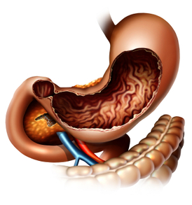
    Stomach cancer is referred to as gastric cancer. The most common type of gastric cancer is called adenocarcinoma. This starts in the glandular tissue that composes the lining of the stomach and accounts for 90% to 95% of all gastric cancers. Other forms of gastric cancer include lymphomas, which involve the lymphatic system and sarcomas, which involve the connective tissue (such as muscle, fat, or blood vessels).

    iStock_000002366269XSmall.jpg

    Generally, the best chance for cure of gastric cancer is when it is found and treated at a very early stage. Unfortunately, the outlook is typically poor if the cancer is already at an advanced stage when discovered.

    Gastric cancer can develop in any part of the stomach and may spread throughout the stomach and to other organs, such as the esophagus, liver and lungs.  Gastric cancer is responsible for about 800,000 deaths worldwide per year.

    The signs/symptoms include indigestion and/or heartburn, loss of appetite, especially for meat, abdominal discomfort or irritation, weakness and fatigue, bloating of the stomach, usually after meals, abdominal pain in the upper abdomen, nausea and occasional vomiting, diarrhra or constipation, weight loss, vomiting, blood in the stool, which will appear as black. This can lead to anemia, and dyspagia (trouble swallowing), which suggests a tumor in the upper protion of the stomach or extension of the gastric tumor into the esophagus.

    The diagnosis is typically made when abnormal tissue seen in a direct visual (gastroscopic) examination is biopsied. This tissue is then examined under a microscope to check for the presence of cancerous cells. A biopsy, with subsequent histological analysis, is the only sure way to confirm the presence of cancer cells.

    The clinical stages of stomach cancer are:

    Stage 0. Limited to the inner lining of the stomach.

    Stage I. Penetration to the second or third layers of the stomach (Stage 1A) or to the second layer and  nearby lymph nodes (Stage 1B).

    Stage II. Penetration to the second layer and more distant lymph nodes, or the third layer and only nearby lymph nodes, or all four layers but not the lymph nodes.

    Stage III. Penetration to the third layer and more distant lymph nodes, or penetration to the fourth layer and either nearby tissues or nearby or more distant lymph nodes.

    Stage IV. Cancer has spread to nearby tissues and more distant lymph nodes, or has metastatized to other organs.

    Treatment for gastric cancer depends on both the tissue type and the stage of the cancer. Treatment for adenocarcinoma may include:

    1. Surgery– the goal of surgery is to remove all of the cancer and allow for a margin of healthy tissue,  when possible. The surgeon also removes lymph nodes surrounding the stomach to determine if they  have been invaded by cancer cells. Removing part of the stomach may relieve signs and symptoms of a  growing tumor in people with advanced stomach cancer. In this case, surgery generally does not cure the  cancer, but it can make the patient more comfortable. This is known as palliative therapy.

    2. Radiation Therapy-uses high-powered beams of energy to kill cancer cells. It can be used before surgery (neoadjuvant radiation) to shrink a stomach tumor so it’s more easily removed. Radiation therapy can also be used after surgery (adjuvant radiation) to kill any cancer cells that might remain.  Radiation is often combined with chemotherapy. In cases of advanced cancer, radiation therapy may be  used to relieve side effects caused by a large tumor. Radiation therapy can cause diarrhea, indigestion,  nausea and vomiting.

    3. Chemotherapy-is a drug treatment that uses chemicals to kill cancer cells. Chemotherapy drugs can  kill cancer cells that may have spread beyond the stomach. Chemotherapy can be given before surgery  (neoadjuvant chemotherapy) to help shrink a tumor so it can be more easily removed. And it can be  given after surgery (adjuvant chemotherapy) to kill any cancer cells that might remain in the body.    Chemotherapy is often combined with radiation therapy. Chemotherapy may be used alone in patients  with advanced stomach cancer to help relieve signs and symptoms. Side effects of chemotherapy depend  on which drugs are used, and which drugs are used depends upon the type of cancer being treated.

    4. Clinical Trials-are always being done at research cancer centers to study new treatments and new ways of using existing treatments. Participating in a clinical trial may give a patient a chance to try the  latest treatments. In some cases, researchers might not be certain of a new treatment’s side effects.

    Depending on the stage at diagnosis, the average 5 year survival rates for adenocarcinoma of the stomach are as follows:

    Stage Ia – 71%

    Stage Ib – 57%

    Stage IIa – 45%

    Stage IIb – 33%

    Stage IIIa – 20%

    Stage IIIb – 14%

    Stage IV – Less than 4%.

    Medical malpractice may be seen when there is a significant and negligent delay in the diagnosis of gastric cancer. As the above shows, if the diagnosis is delayed, and the stage advances, the prognosis for 5 year survival rates becomes poor.  During patient screening, a doctor should be aware of the risk factors for gastric cancer that include smoking cigarettes, a diet high in salted, smoked, pickled meats or foods, stomach inflammation, a history of Helicobacter pylori infection, gastric polyps, pernicious anemia, and a family history of one or more relatives diagnosed with gastric cancer.  Screening for one type of adenocarcinoma called HDGC or Hereditary Diffuse Gastric Cancer includes genetic testing for the CDH1 gene mutation.  In families with the CDH1 gene mutation, intense surveillance may be appropriate and in some cases prophylactic gastrectomy.

    Aside from upper endoscopy, other helpful diagnostic tests for gastric cancers include a double contrast barium x-ray, ultrasound, or a CT scan; however, a negative barium x-ray study alone may give false assurance and is sometimes a reason for a delay in diagnosis. Research suggests that improvement in five-year survival requires not only improved awareness by patients, but especially improved diagnostic methods and screening programs by their doctors.


  4. Hydrocephalus and Medical Malpractice

    Posted by Dr. Jack Sacks, Esq.on February 16, 2016

    Hydrocephalus is also known as “water in the brain.”  It is a condition in which there is an abnormal accumulation of cerebrospinal fluid (CSF) in the ventricles, which are cavities in the brain.  In infants with hydrocephalus, CSF builds up in the central nervous system, causing the soft spot near the front of the scalp, known as the anterior fontanelle, to bulge and the head to be larger than expected. There is also a soft spot near the back of the head known as the posterior fontanelle. Early symptoms may include sundowning, which is when the eyes appear to gaze downward; irritability; seizures; separated scalp sutures; sleepiness; and vomiting.

    In newborns with hydrocephalus, the head circumference rapidly enlarges and may surpass the 97th percentile when measured at pediatric visits.  If the skull bones have not yet firmly joined together at this age, bulging, firm anterior and posterior fontanelles may be present when the patient is in an upright position. The infant may exhibit fretfulness, poor feeding, and frequent vomiting.

    As the hydrocephalus progresses, torpor, a state of sluggishness and reduced metabolism, sets in and the infant shows lack of interest in his/her surroundings. In a later stage, the upper eyelids become retracted and the eyes turn downwards (due to hydrocephalic pressure on the mesencephalic tegmentum and paralysis of upward gaze). Movements become weak and the arms may become tremulous.  There may be reduction of vision. Over time, the head becomes so enlarged that the growing child becomes bedridden and he/she is unable to lift or even move this huge object attached to its neck.

    Hydrocephalus is diagnosed through clinical neurological evaluation and by using cranial imaging techniques such as ultrasonography, computed tomography (CT), magnetic resonance imaging (MRI), or pressure-monitoring techniques. The most common initial diagnostic test to determine hydrocephalus is an image of the brain (CT Scan or MRI) to identify the enlarged ventricles (spaces) within the brain that are typical of hydrocephalus.

    Hydrocephalus is often treated with the surgical placement of a shunt system. This system diverts the flow of CSF from a site within the central nervous system (CNS) to another area of the body where it can be absorbed.  A shunt is a flexible but sturdy tube composed of silicone and plastic.   A shunt system consists of a shunt tube, a catheter, and a valve. One end of the catheter is placed into one of the dilated a ventricles inside the brain.  Depending on the situation, it could also be placed into a cyst or somewhere near the spinal cord. The other end of the catheter is usually placed within the abdominal cavity.  It could also be placed into any area, such as the heart, where the extra cerebrospinal fluid can be drained and absorbed.  The valve located along the catheter maintains one-way flow and regulates the rate of CSF flow.

    Hydrocephalus can be effectively treated with a shunt; however, there is still a potential for complications. The shunt can become blocked. Symptoms of blockage include headache and vomiting. There may be other problems with the shunt such as kinking, tube separation, or infection in the area of the shunt. Other complications may include infections such as meningitis or encephalitis, intellectual impairment, and nerve damage.  Untreated hydrocephalus has a 50-60% death rate. Survivors have varying degrees of intellectual, physical, and neurological disabilities.


  5. Spinal Cord Compression and Cauda Equina Syndrome

    Posted by Dr. Jack Sacks, Esq.on January 03, 2016

    There are approximately 12,000 new cases of spinal cord compression annually in the United States with the average age of victims being 39.5 years old. Causes of spinal cord compression include trauma (such as auto accidents, falls, sports injury, epidural injection), spinal abscess, tumor, hematoma or blood clot, ruptured or herniated disk, and spinal stenosis. . Early symptoms may include the start of loss of movement or feeling in the arms or legs, back pain, and the loss of bowel or bladder function or control. Spinal cord compression of sudden onset constitutes a medical emergency, as the longer the duration of symptoms before cord pressure relief is obtained by surgical intervention (laminectomy), the greater the chance of permanent injury. Diagnosis is by clinical exam and other testing such as X-ray, CT, and MRI. MRI is usually the most accurate study to detect spinal cord compression as details of both bony and soft tissue abnormalities in the spinal column may be visualized. MRI may also be the preferred study if spinal cord injury occurs during pregnancy as MRI offers reduced radiation exposure to the fetus. However, after an accident, traction devices to immobilize the spine and life support equipment may preclude the use of MRI.

    The spinal column is comprised in descending order of the cervical, thoracic, lumbar, and sacral areas. The actual spinal cord ends at the level of L-1. Below the level of L-1, the cord branches into a bundle of spinal nerve roots from L-1 through L-5 and S-1 through S-5 that resembles a horses tail referred to as the cauda equina. When the nerve roots of the cauda equina are compressed permanent neurogenic injury may occur. This may be referred to as cauda equina syndrome (CES). Causes of compression of these nerve roots include the same types of causes as listed above for spinal cord compression. Symptoms of early onset of cauda equina syndrome include numbness in the groin (saddle anesthesia), loss of bowel or bladder function or control, weakness in the legs, and absence of ankle reflexes. Early diagnosis and treatment of CES is important as the longer the duration of symptoms, the more likely permanent neurological injury will occur such as paralysis and incontinence. Cauda equina syndrome of sudden onset is a medical emergency and treatment generally involves surgical decompression of the affected nerve roots. A laminectomy may be performed to relieve pressure on the nerve root in cases where there is a herniated or ruptured disk, hematoma, abscess, or tumor.

    In spinal cord and nerve root compression cases involving sudden onset of neurological symptoms, medical malpractice may arise when there has been an unreasonable delay in diagnosis and prompt surgical intervention by the doctors or hospital. Allegations of medical malpractice may also include a failure to diagnose and treat a vertebral fracture of the neck or back that required traction or immobilization which was not performed, thereby allowing pathological movement that resulted in spinal cord compression.


  6. Leg Amputation and Medical Malpractice

    Posted by Dr. Jack Sacks, Esq.on January 02, 2016

    Amputation is the surgical removal of all or part of an extremity. The most common amputation surgery is above or below the knee.  The indications for leg amputation include severe trauma, significant tumor in the bone or muscle, lack of blood circulation due to peripheral arterial disease, worsening or uncontrollable infection, failed management of acute compartment syndrome, failed management of Charcot’s degenerative osteoarthropathy, or debilitating extremity paralysis from infection or pressure-related complications. Failute to timely diagnose and treat infection, tumor, pressure sores, vascular disease, compartment syndrome, and Charcot’s all may result in the unneccessary amputation of a leg and give rise to allegations of medical malpractice against negligent health care practitioners.

    iStock_000026640818XSmall.jpg

    The amputation procedure varies depending on the extremity undergoing the operation. To determine the operation site and the amount of tissue to remove, the surgeon relies on the following factors: the patient’s pulse, skin temperature, areas of reddened skin, and sensitivity to touch in the affected extremity. The presence of a palpable pulse proximal to the level of amputation is a positive predictor for successful healing; however, the absence of a pulse does not necessarily reflect future wound healing failure. The level of the amputation is based on the extent of the damaged tissue, the healing potential of the area, and the rehabilitation potential of the patient. In addition to a thorough clinical examination, objective tests such as ankle pressures, toe pressures, transcutaneous oxygen measurements, and skin perfusion pressures are useful.

    The preoperative evaluation and preparation involves medical risk assessment, nutrition assessment, prosthetic and rehab consultation, and possibly a psychological consultation. The diseased tissue is removed along with any crushed bone and the maximal amount of healthy tissue is left behind. The blood vessels and nerves in the surrounding area are sealed off. Following the amputation, the site can be left open due the possibility of further amputation or covered with skin flaps and closed. The remaining muscles in the area are shaped so the end of the limb can be fitted for prosthesis, also known as an artificial limb.

    Thromboprophylaxis is recommended for all patients undergoing major lower extremity amputation because patients are at high risk for thromboembolism, the blocking of a blood vessel by a particle that has separated from a blood clot at the formation site. Antibiotic prophylaxis is typically recommended within one hour of skin incision for lower extremity amputation due to high risk for surgical site infection.

    Generally, the patient undergoes physical rehabilitation soon after surgery and practice with the prosthesis can begin 10-14 days after surgery. The patient’s postoperative outcome is dependent upon preoperative functional status, comorbidities, and the level of amputation. Wound healing must be monitored and dressing changes performed. Patients with advanced diabetes, significant heart disease, or serious infection are at a greater risk of complications from the procedure. Possible complications include infection, joint contracture, necrosis, deep vein thrombosis, pulmonary embolism, hematoma, and wound opening. In addition, patients may experience phantom pain, a sense of pain in the amputated limb described as burning aching, or electric. Other causes of pain such as ischemia, infection, neuroma, or pressure related wounds should be excluded before determining the diagnosis as phantom pain.


  7. Appendicitis: Early Diagnosis and Treatment are Essential

    Posted by Dr. Jack Sacks, Esq.on December 01, 2015

    Appendicitis is a medical emergency that requires immediate surgery to remove the appendix.  If left untreated, an inflamed appendix will eventually rupture.  When this happens intestinal contents (stool and bacteria) spill into the abdominal cavity causing infectious peritonitis, a serious and toxic inflammation of the abdominal cavity’s lining (the peritoneum).  This condition can be fatal unless it is treated quickly with surgery and strong antibiotics.

    SYMPTOMS: Usually pain is the first symptom, starting in the mid abdomen around the navel, and except in children below 3 years old, the pain tends to localize in the right lower quadrant within a few hours.  The abdominal wall becomes sensitive to gentle pressure, and the pain can be elicited through various tests the physician will use to bring it out.  One such sign is when the abdomen is gently pushed down and quickly released.  This is known as rebound tenderness and is a clinical sign the peritoneum is inflamed.  If the appendix is located beneath the cecum (first part of the colon), it may fail to elicit tenderness (silent appendix).  And if the appendix lies entirely within the pelvis, the region below the addomen, there could be a complete absence of the abdominal signs and symptoms. In such cases, a digital rectal exam will cause discomfort localized to the region of the appendix in the right lower quadrant.  Also, if the abdomen on palpation is rigid, which is known as involuntary guarding, there should be a strong suspicion of peritonitis requiring urgent surgical intervention.  The physician can perform certain other maneuvers, such as bending and rotating the right hip, and extending the hip in the prone position, which will bring about pain consistent with inflammation caused by appendicitis.  The next symptoms usually experienced are naussea and vomiting, as well as constipation.  Eventually as the inflammation progresses, fever will occur.

    BLOOD TESTS: When appendicitis is suspected, blood tests such as a CBC need to be done to try to confirm the diagnosis.  More than 80% of adults with appendicitis have a white blood cell count greater than 10,500 cells/mm3.  Another blood test commonly used is the determination of C-reactive protein (CRP),an acute-phase reactant synthesized by the liver in response to infection or inflammation.

    DIAGNOSTIC IMAGING: The plain film of the abdomen, known as a KUB is typically taken. Visualization of an appendicolith (a white colored defect in the right lower quadrant) in a patient with symptoms consistent with appendicitis is highly suggestive of appendicitis, but this occurs in fewer than 10% of cases. Another X-ray that may be utilized is the single-contrast Barium enema, which can be performed on an unprepared bowel. Absent or incomplete filling of the appendix with contrast barium coupled with pressure effect or spasm in the cecum suggests appendicitis. Though cheap in cost, once thick barium contrast is instilled, other more definitive tests will not be able to be performed.

    The appendix may be evaluated via transabdominal sonography. Many physicians believe that ultrasonography should be the initial imaging test in pregnant women and in children due to its safety. Abdominal CT scanning has become the most important imaging study in the evaluation of patients with appendicitis.  Advantages of CT scanning include its superior accuracy when compared with other imaging techniques, as well as its ready availability, noninvasiveness, and potential to reveal alternative diagnoses. MRI plays a relatively limited role in the evaluation of appendicitis due to high cost, long scan times, and limited availability.  The lack of dangerous radiation exposure makes it an attractive modality in pregnant patients where ultrasound is not helpful in making a diagnosis.

    In today’s day and age, the diagnosis of appendicitis should generally not be missed by an emergency room physician.  If in doubt a surgeon should be called to evaluate and remove the appendix, which can be done laparoscopically, so long as it remains locally inflamed or walled off, and before there is free perforation into the abdominal cavity.  A delay in diagnosis can result in lifelong complications affecting bowel function due to the scarring of the peritoneal lining.


  8. BREAST CANCER AND MEDICAL MALPRACTICE

    Posted by Dr. Jack Sacks, Esq.on October 18, 2015

    Breast cancer affects millions of women in America and some studies indicate up to one out of eight women will be diagnosed with breast cancer over the course of her lifetime. The survival rate of breast cancer that is detected and treated early is much better than when a malignant tumor is found after it hasalready spread to lymph nodes or surrounding tissue.  Therefore, it is important that every effort be made to diagnose and treat breast lesions as quickly as possible.iStock_000013080813XSmall-thumb-300x400-8870-thumb-300x400-8871-thumb-300x400-8872.jpg

    One of the mainstays in breast cancer detection is mammography. A mammogram involves an X-ray taken of the breasts, usually taken from several angles. Some abnormalities on mammograms such as masses or calcifications can alert the healthcare provider and patient that additional diagnostic testing needs to be done. The mammogram is one of the main methods of screening for breast cancer. Other imaging methods are less commonly used for screening, such as sonography (ultrasound), and MRI. However, these diagnostic methods can be used to add to information already obtained from a mammogram study when a patient is evaluated and uncertainty or an abnormality exists.

    Patients need to be informed about the need for breast cancer screening. The mammogram, ultrasound, or MRI tests need to be read and interpreted correctly in order to diagnose problems and treat them early. In the case of breast cancer, early detection and diagnosis is key to improved cure rates.  Special circumstances and risk factors (such as family history) may point to additional diagnostic testing to help identify breast cancer in those women at high risk. If tests are not interpreted correctly, or the results are not acted on in a timely or proper fashion by the healthcare provider, medical malpractice may be the result.

    Sometimes patients or healthcare providers feel a breast mass or lump on examination. This often requires further evaluation usually beyond just a “regular” screening mammogram. As noted above, additional mammogram views, ultrasound, and/or MRI can help characterize the type of mass. However, breast masses usually warrant a visit to a surgeon or breast specialist to evaluate the patient for biopsy. Breast biopsies can be much less invasive to patients than in years past. During a “stereotactic” breast biopsy, the doctor employs mammography to locate the specific abnormal area and then a special instrument is used to remove cells from that precise area. Ultrasound can also be used to identify an abnormal area that is then biopsied. These methods are particularly useful when a breast mass cannot be felt. Sometimes when a mass can be felt, or palpated, the biopsy can be done without using X-ray or ultrasound localization.

    Breast cancer screening can help identify women with breast cancer, as well as pre-cancerous lesions and benign breast conditions. Medical malpractice may occur as a consequence of failure to adequately evaluate, identify, screen, or diagnose breast cancer. Allegations of medical malpractice may include a failure by the internist to refer the patient to a breast specialist or surgeon for evaluation, failure by the radiologist to properly read a mammogram, ultrasound, or MRI, and/or failure by the pathologist to detect cancer in a breast biopsy that is indeed malignant. With improved methods of screening, diagnosis, and treatment of breast cancer, survival rates should continue to improve.


  9. Persistent Pulmonary Hypertension of the Newborn

    Posted by Dr. Jack Sacks, Esq.on September 09, 2015

    Persistent pulmonary hypertension of the newborn (PPHN) can occur in babies when the circulatory system does not adapt to conditions outside the mother’s uterus. Before birth, the baby’s oxygen needs are met through the placenta in the mother’s uterus. After birth, the baby’s lungs take over this function. After being born, the baby inflates its lungs with its first breaths. Then, a series of changes take place to decrease resistance to blood flowing through the newborn’s lungs. This allows the lungs to take over getting oxygen for the baby’s vital functions.

    Sometimes, the changes that allow blood to preferentially flow from the baby’s heart to the lungs do not proceed normally, or at all. This can occur in approximately 2 out of 1000 births.

    Some PPHN cases are due to problems in the baby’s lung tissue. For example, if baby has damaged lungs from inhaling meconium, or pulmonary damage from prematurity, this can contribute to pulmonary hypertension. Development problems or defects in lung and related blood vessels can also lead to PPHN. However, PPHN can develop even when the lung tissue and blood vessels are normal.

    Some cases of persistent pulmonary hypertension of the newborn have been linked to medications used by pregnant women. Antidepressants such as Zoloft (sertraline), Paxil (paroxetine), Prozac (fluoxetine), Lexapro (escitalopram) and Celexa (citalopram) may be associated with PPHN. The FDA issued a public health advisory in 2006 about the use of these medications in pregnant women.

    It is crucial for pregnant women to review their medical and medication histories with their doctors. Certain medications may need to be adjusted, stopped, or continued according to the patient’s needs and risks to the unborn baby. If potentially damaging medications were used and a birth defect such as PPHN occurred, the victims may have legal rights that need to be protected.


  10. Diabetes and Medical Malpractice: Elevated Blood Glucose or Hypoglycemic Events

    Posted by Dr. Jack Sacks, Esq.on June 02, 2015

    Diabetes affects over 23 million people in the United States and is the 7th leading cause of death.  Uncontrolled high levels of blood sugar can cause vascular damage to the retina of the eye, the filtering system of the kidney, and the peripheral nerves.  In fact, diabetes is the leading cause of adult blindness, kidney failure, and non-traumatic amputations in the United States.  Failure by a health care provider to diagnose, treat, and educate diabetic patients may constitute medical malpractice should a serious injury occur.  Preventative measures such as annual dilated pupil exams,  annual foot exams, and prescription Thumbnail image for iStock_000004641088Large.jpgmedications to control high blood pressure can greatly reduce the risk of injury to the diabetic patient.  Outpatient laser treatment to seal off abnormal blood vessels in the retina may significantly delay or prevent the onset of symptomatic diabetic retinopathy and blindness.  ACE inhibitors are drugs that can lower blood pressure and prevent damage to the filtering system of the kidney.  Effective patient education (by a diabetic educator) about the importance of a maintaining a healthy diet, daily exercise, and proper hygiene are essential to help prevent diabetic complications.

    Diabetes may arise for the first time during pregnancy and cause serious complications.  This type of diabetes is referred to as gestational diabetes and can cause the fetus to become oversized and fat due to the excess glucose in the umbilical blood.  Unfortunately, an oversized fetus may render a vaginal delivery high risk as birth injuries such as shoulder dystocia are more likely to occur.  Failure to diagnose and treat gestational diabetes may result in life threatening complications to the fetus and the mother and may give rise to a medical malpractice claim.

    On the flip side, low blood sugar can cause hypoglycemic episodes that may result in brain damage or death.  Over administration of insulin, or a failure to monitor low blood sugar levels in diabetic patients may constitute medical malpractice.  Reduced blood sugar levels (blood glucose below 65mg/dL) may result in neurogylopenic effects such as impaired judgment, dizziness, slurred speech, seizures, and coma.  In a hospital or nursing home setting, glucose fingerstick testing may be necessary every few hours to prevent a serious hypoglycemic episode from arising.