Medical Malpractice Attorneys
  • Toll Free 1-800-529-9120
  1. Sigmoid Colon Volvulus and Medical Malpractice

    Posted by Dr. Jack Sacks, Esq.on February 03, 2012

    The term volvulus is derived from the Latin word volve, which means to twist. A colonic volvulus occurs when a part of the colon twists on its mesentery, resulting in acute, subacute, or chronic colonic obstruction.  Volvulus involving the sigmoid colon is the most common, occuring in 75% of cases.  Sigmoid volvulus occurs when the last part of the large bowel just before the rectum (named for its “S” shape) twists on its self.  It is common in elderly men, but it is likely to occur in anyone with a redundant sigmoid colon.

    More than 60-70% of patients present with acute symptoms; the remainder present with subacute or chronic symptoms. A history of chronic constipation is common. The patient may describe previous episodes of abdominal pain, distension, and obstipation suggestive of repeated, subclinical episodes of volvulus.  With continued obstruction, nausea and vomiting can occur. The development of constant abdominal pain is ominous. It indicates there may be a closed loop obstruction with significant intraluminal pressure, which can lead to ischemic gangrene and bowel wall perforation.

    Abdominal distension is generally massive and characteristically tympanitic over the gas-filled, thin-walled bowel loop. The presence of overlying or rebound tenderness raises the concern of peritonitis due to ischemic or perforated bowel. A patient with a history of acute volvulus episodes that spontaneously resolve can experience marked distention with minimal abdominal pain.

    A radiographic film of the abdominal will demonstrate a huge air filled distended bowel frequently in the shape of an inverted “U,” with the convexity of the “U” facing the right upper abdominal quadrant.  A barium enema will show dilation in the sigmoid colon due to a twist. A physician may refer to an area of complete obstruction with some twisting as the “bird beak” sign.

    CT scans can demonstrate crossing sigmoid transitions, tagged the X-marks-the-spot sign, and folding of the sigmoid wall by partial twisting, called the split-wall sign.  However, the most sensitive finding on CT is a sigmoid colon transition point, which is seen in 95% of scans, and a disproportionate enlargement of the sigmoid colon, noted in 86% of cases.

    Colonoscopy or flexible sigmoidoscopy could be done to both confirm the diagnosis as well as attempt to treat the obstruction. Barium enemas can also reduce the obstruction when the pressure of the fluid rushing into the bowel unwinds it.

    For treatment, the first step is to free the acute obstruction, and then to fix the redundant part of the bowel to prevent reoccurrence.  In up to 90% of patients with sigmoid volvulus, the condition recurs after untwisting with methods as noted above. For this reason, anyone with a sigmoid volvulus needs to undergo an operation during the same admission to either remove or fix down the excessive bowel length.

    Once the diagnosis of sigmoid volvulus is confirmed, treatment must be immediate. A delay in treatment represents a greater likelihood of bowel wall death and gangrene. Up to 80% of people with this condition die from gangrene if intervention is delayed. In the event this diagnosis is missed and bodily injury results, the treating health care provider is at risk of a medical malpractice lawsuit.

  2. Triple Negative Breast Cancer and Medical Malpractice

    Posted by Dr. Jack Sacks, Esq.on January 31, 2012

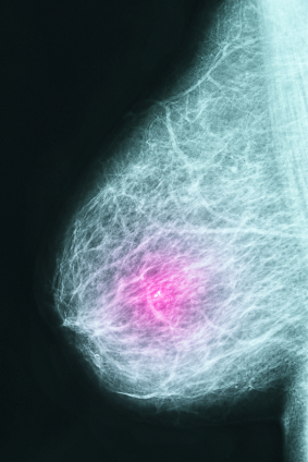
    By dividing breast cancer into molecular breast cancer subtypes, physicians and researchers can devise better approaches and treatments for dealing with the disease. Most studies divide breast cancer into four major molecular subtypes: Luminal A, Luminal B, Triple-negative, and HER2/neu. Triple-negative breast cancer is characterized by tumors that lack estrogen, progesterone, and human epidermal growth factor receptors.

    iStock_000018505784XSmall.jpg

    Higher rates of triple negative breast cancer have been recorded in younger women, Hispanic and African American women, and women of lower socioeconomic class. Triple negative cancer tends to have a poorer prognosis than other types of breast cancer that have more available and effective treatments.

    Studies have demonstrated that triple-negative breast cancer possesses a higher probability for recurrence and spreading beyond the breast. This risk is greater within the first few years post-treatment but over time the risk becomes similar to other types of breast cancer. In addition, triple-negative tends to present as a higher grade cancer, meaning that a greater number of cancer cells are abnormal in appearance.

    Although surgical and radiation treatments are similar for different types of breast cancer, drug treatments usually vary. Many therapies for breast cancer target cellular receptors. A receptor is a molecule generally located on the surface of cells that is involved with chemical signaling. When a molecule binds to a receptor, it causes the cell to perform certain actions such as division or multiplication. Targeted drug treatments like tamoxifen or Herceptin, designed to treat HER2 positive breast cancer, have not been developed for triple negative. A patient with triple-negative does not express estrogen, progesterone, and HER2/neu receptors so therapy against these receptors is ineffective.

    Standard treatment for triple-negative breast cancer is chemotherapy. Current treatments include anthracylines, taxanes, ixabepiplone, platinum agents, and biologic agents. Patients undergoing chemotherapy can experience side effects such as vomiting, nausea, alopecia, mucositis, myelosuppression, etc. Surgery and radiation are additional avenues of treatment. Currently, cancer experts are studying several promising drug strategies targeted for triple negative breast cancer such as PARP inhibitors.


  3. Septic Arthritis and Medical Malpractice

    Posted by Dr. Jack Sacks, Esq.on December 31, 2011

    Septic arthritis, also known as reactive arthritis and bacterial arthritis, is inflammation of a joint caused by infection. When it develops, it represents a medical emergency. It develops when bacteria or fungus is transmitted through an individual’s bloodstream to a joint. Individuals can develop septic arthritis at any age but it is less common between the ages of three to adolescence.

    The majority of acute cases involve bacteria such as staphylococcus or streptococcus. However, children who develop septic arthritis tend to be infected with Group B streptococcus or Haemophilus influenza, if they have not been vaccinated. The number of cases that progress to a chronic infection are less common. Chronic infections are generally caused by organisms such as Mycobacterium tuberculosis and Candida albicans.

    Overall, the most common sites of infection are the hip and knee joints. The following are considered risk factors for septic arthritis: artificial joint implants, bacterial infection, chronic disease (diabetes, rheumatoid arthritis, and sickle cell disease), intravenous drug use, immunosuppressive medications, and recent joint surgery or arthroscopy.

    Septic arthritis generally presents with a rapid onset of symptoms. Patients develop a fever in addition to joint swelling, redness, and pain in the infected joint. Patients may also experience pseudoparalysis, which is an inability to move the extremity possessing the infected joint. To diagnose septic arthritis, physicians will aspirate the joint fluid to check the cell count, look for crystals under the microscope, and perform a gram stain and culture. In addition, other tests include a blood culture and an X-ray of the affected joint.

    Possible complications include joint degeneration and permanent joint damage, which can occur if appropriate treatment is delayed. If antibiotics are promptly given to treat the infection the patient has a good prognosis. If fluid accumulates in the joint, aspiration may be necessary. Aspiration involves a needle being inserted into the joint to drain off the excess fluid. Only severe cases require surgery to remedy the infected joint fluid; however, if a prosthetic joint is affected, it may need to be replaced.


  4. Pituitary Adenomas and Medical Malpractice

    Posted by Dr. Jack Sacks, Esq.on October 25, 2011

    A pituitary adenoma is generally a benign, slow growing tumor that occurs in the pituitary gland. The pituitary gland is a small, bean shaped structure that lies at the base of the brain. It has a central role in the regulation of hormones that affect the body such as Adrenocorticotropic hormone (ACTH), Growth hormone (GH), Prolactin, and Thyroid-stimulating hormone (TSH).

    Approximately 1 in 1,000 individuals have pituitary adenomas. They are generally not cancerous but may invade nearby structures.  They are classified based on size. A microadenoma is less than 1 cm in diameter whereas a macroadenoma is larger than 1 cm in size.

    iStock_000017548218XSmall.jpg

    Based on whether the pituitary adenoma is a hormone-producing or hormone-inactive tumor, the patient will present with different symptoms. Hormone-producing tumors will make excessive amounts of an active hormone so symptoms present as a hormonal imbalance. The three most common hormone-producing adenomas are Prolactinomas, Growth hormone-secreting pituitary adenoma, and ACTH-secreting pituitary adenoma.

    If the patient has a large hormone-inactive or hormone-producing tumor, it may compress surrounding brain structures due to its size.  Large pituitary tumors may compress the pituitary gland contributing to pituitary failure, which can lead to sexual dysfunction, inadequate body cortisol levels, and hypothyroidism. Other possible presentations relating to compression of brain structures include visual loss, headache, the “stalk effect”, and pituitary apoplexy.  The “stalk effect” is due to the compression of the pituitary stalk, the structure connecting the brain to the pituitary gland, which leads to a mild elevation in the hormone prolactin. Higher levels of prolactin in females can contribute to irregular menstrual cycles. Pituitary apoplexy presents with an abrupt headache and visual loss. It can occur under two different situations. The pituitary adenoma can bleed internally causing a sudden increase in size or the tumor can outgrow its blood supply and the dead tissue will swell.

    Pituitary adenomas can be diagnosed based on endocrine function testing, imaging, and visual field testing. Often times, visual problems may be the only symptom present.  Optometrists and ophthalmologists should seriously consider the possibility of a pituitary tumor when the patient presents with an unexplained loss of visual field (especially peripheral vision), double vision, or blurred vision as the growth of a pituitary tumor can cause compression on the visual pathway (retina, optic nerve, etc). A delay in diagnosis of a pituitary adenoma may result in permanent loss of vision due to the damage caused by this compression. Endocrine function testing evaluates cortisol, follicle-stimulating hormone, lutenizing hormone, insulin growth factor-1, prolactin, testosterone/estradiol, and thyroid hormone levels. The preferred method of imaging is MRI, which screens for adenomas larger than 4 mm.

    Treatment for pituitary adenomas depends on the presence of hormone production, size of the tumor, invasion of the tumor into surrounding structures, and the age and health of the patient. Drug therapy is used to treat hormone-producing tumors. For example, bromocriptine and cabergoline are used to treat tumors secreting prolactin because these medications decrease prolactin levels and tumor size. Pituitary adenomas that require surgery are usually minimally invasive techniques, where the tumor is removed through the nose.  On the other hand, radiation therapy involves high doses of radiation being delivered to the tumor. It is a treatment utilized for pituitary adenomas that cannot be controlled by drug therapy or surgical intervention. Patients have the best outlook when the entire tumor can be removed.


  5. Lumbar Puncture – Indications and Usage

    Posted by Dr. Jack Sacks, Esq.on October 07, 2011

    Lumbar puncture is a procedure performed in the lower back area, where a needle is inserted between two vertebrae to remove a sample of cerebrospinal fluid (CSF). The patient flexes his/her back to widen the spaces between the vertebrae so it is easier for the physician to access the region. The back is washed with antiseptic soap or iodine and covered with a sterile sheet. A local anesthetic is used to numb the area and then a thin hollow needle is inserted through the spinal membrane and into the spinal canal. During this portion of the procedure, the patient generally feels pressure. The CSF pressure is measured, a small amount of fluid is removed, and the pressure is measured again. The needle is removed and the site is bandaged. The total procedure takes about 45 minutes.

    Thumbnail image for Thumbnail image for iStock_000016020161XSmall.jpg

    Lumbar puncture is used to collect CSF for analysis to help in diagnosing conditions such as subarachnoid hemorrhage (typically caused by ruptured aneurysm or traumatic brain injury), meningitis (inflammation of the membranes around the brain), and cancers of the brain or spinal cord. Lumbar Puncture may also be utilized to inject anesthetic medications, chemotherapeutic drugs, contrast material, or radioactive substances into CSF.

    Lumbar Puncture may be used to identify increased or decreased CSF pressure.  Increased CSF pressure can be caused by increased intracranial pressure as is seen with traumatic brain injury, ruptured aneurysm, and sometimes hydrocephalus. On the other hand, decreased CSF pressure can be caused by spinal cord tumor, shock, fainting, or diabetic coma.  Normal CSF appears clear and colorless. When an infection is present, the CSF may look cloudy and be yellow or pink in color. Infection may be suspected if there is an increased level of white blood cells and/or protein. Increased CSF glucose indicates hyperglycemia whereas decreased CSF glucose may reflect hypoglycemia, bacterial or fungal infection, tuberculosis, or meningitis. If tumor cells are detected, the patient may have cancer in the brain, spinal cord, or CSF.  If there are increased gamma globulin levels, the patient may be suffering from multiple sclerosis, neurosyphilis, or Guillan-Barre syndrome. The analysis of the CSF by the lab and the measuring of CSF pressure recorded from the lumbar puncture help in determining a diagnosis in many serious clinical situations.


  6. Hemochromatosis and Medical Malpractice

    Posted by Dr. Jack Sacks, Esq.on August 26, 2011

    Primary Hemochromatosis occurs when too much iron builds up in the body. It is usually caused by a specific genetic problem that causes too much iron to be absorbed. If there is too much iron in the diet, the extra iron is absorbed in the gastrointestinal tract and builds up in the body tissues, particularly the liver, heart and pancreas. The result is damage to these organs. Primary hemochromatosis is the most common genetic disorder in the United States, affecting an estimated 1 of every 200 to 300 Americans.

    Hemochromatosis affects more men than women. It is particularly common in Caucasians of western European descent. Symptoms are often seen in men between the ages of 30 and 50 and in women over 50, although some people may develop problems by age 20. There is increased risk if a relative has or had the condition.

    Symptoms include severe fatigue (74%), impotence (45%), and arthralgia (44%), abdominal pain, decreased sex drive, lack of menstruation in women, hypothyroidism, and bronzing of the skin. Clinical manifestations include liver disease, skin pigmentation, diabetes mellitus joint damage, (arthropathy), impotence in males, and cardiac enlargement, with or without heart failure or conduction defects. The most common presentation is a patient with an enlarged liver (hepatomegaly), skin pigmentation, and arthritis.

    Thumbnail image for iStock_000005267204XSmall.jpg

    Along with an elevated serum iron level, other abnormal lab values include an elevated transferin saturation, which measures the amount of iron bound to transferrin, a protein that transports iron, an elevated ferritin, a test used to evaluate the body’s iron stores, and an abnormal liver profile (a group of blood tests used to evaluate liver function). Commonly alanine amino transferase (ALT) or alkaline phosphatase are elevated.  When iron stores increase in the pancreas, pancreatitis leading to potentially fatal diabetes can occur. Iron overload damages the liver causing jaundice, which leads to fatal hepatic cirrhosis. The heart is another organ that excess iron can damage, causing it to fail.

    Fortunately, primary hemochromatosis has a relatively simple treatment, and these complications, if caught early enough, can usually be reversed by treatment of the iron overload causing them. This treatment in an otherwise-healthy person consists of regularly scheduled phlebotomies (bloodletting). When first diagnosed, the phlebotomies may need to be performed fairly frequently, perhaps as often as once a week, until iron levels can be brought to within the normal range. Once iron and other markers are within the normal range, phlebotomies may be scheduled every other month or every three months depending upon the patient’s rate of iron absorbtion.

    For those patients unable to tolerate routine bloodletting, or whose blood count is not high enough to allow for it, there is a chelating agent, known as desferoxamine (deferoxamine) that can be used. Chelating agents such as desferoxamine bind with iron in the bloodstream and enhances its elimination from the organs involved via the urine and feces. Typical treatment for chronic iron overload requires subcutaneous injection over a period of 8–12 hours daily.

    Primary hemochromatosis must be considered in any patient presenting with an enlarged liver. It also must be suspected if any one liver blood test, seen on any routine metabolic profile, is elevated without an explanation. The same is true for elevated fasting blood sugar levels. This is a disorder that can be successfully treated. And if caught early, a patient can expect to live a normal lifespan. Once the heart, pancreas or liver are damaged, life expectancy is considerably diminished.  Failure by a doctor to timely diagnose hemochromatosis before permanent damage to any bodily organ occurs, may constitute medical malpractice.


  7. Premature Birth May Result in Injury to the Newborn

    Posted by Dr. Jack Sacks, Esq.on August 09, 2011

    Premature birth can often result in complications for the newborn. Preterm or premature birth refers to babies born before 37 weeks of a normal 40 week long pregnancy.

    Some complications that can affect babies born prematurely are lung complications from lack of pulmonary maturity or development. Eye problems can result in vision difficulties or blindness. Premature babies may be more prone to infections and intestinal problems. They also are at increased risk for bleeding in the brain which can have devastating effects. Conditions ranging from learning disabilities to cerebral palsy and seizures are more common in babies born prematurely.

    Thus it is extremely important that premature labor be diagnosed in a timely fashion by the patient’s health care provider. Steps can be taken to treat the underlying cause of the preterm labor. For example, if a maternal infection is present that is related to the preterm contractions , the infection needs to be diagnosed and treated. Certain conditions, such as twins, increase the risk of premature labor. Also, some anatomic abnormalities of the cervix or uterus can elevate the risk as well.

    Although it may not be possible to prevent premature birth, certain steps can be taken to prolong the time until delivery. This may give time for medications to be administered that may significantly improve the newborns outcome by enhancing lung development or decreasing the risk of intestinal problems. Infections that are properly diagnosed and treated may likewise improve the premature baby’s situation.

    Medical malpractice may occur when a pregnant patient’s symptoms, such as premature contractions, bleeding, or rupture of membranes are not properly evaluated and managed. If needed interventions such as medication to improve lung function, or antibiotics, are not given in cases where they are indicated, malpractice may have occurred. An evaluation by attorneys with experience and knowledge of these complex medical and legal issues can help clarify issues for victims of medical malpractice.


  8. Ventricular Septal Defect in the Newborn

    Posted by Dr. Jack Sacks, Esq.on May 24, 2011

    A ventricular septal defect (VSD) refers to a hole in the ventricular septum, the wall dividing the left and right ventricles of the heart. VSDs are the most common congenital cardiac anomalies. They are found in30-60% of all newborns with a congenital heart defect, or about 2-6 per 1000 births. During heart formation, when the heart begins life as a hollow tube, it begins to partition, forming a septa. If this does not occur properly it can lead to an opening being left within the ventricular septum.

    As noted above, during ventricular contraction some of the blood from the left ventricle is forced into the right ventricle from where it enters the lungs through the pulmonary arteries and then reenters the left ventricle via the pulmonary veins and left atrium.  This refluxing of blood causes volume overload on the left ventricle.  This increased blood volume also increases pressure in the right venticle eventually causing pulmonary hypertension with its associated symptoms. This effect is more noticeable in those with larger defects, who may present with shortness of breath, poor feeding and failure to thrive in infancy.

    Because structural cardiac anomalies are estimated to occur in 8 of every 1,000 live births, it is imperative they be discovered before birth. Cardiovascular anomalies, such as VSD, are frequently associated with other congenital anomalies because the heart is among the last organs to develop completely in the embryo. The guidelines for routine prenatal evaluation of both the American College of Radiology and the American Institute of Ultrasound in Medicine require evaluation of the fetal heart.  To not do so, may be consistent with medical negligence or medical malpractice.

    The ultrasound view that is most commonly used is the four-chamber view of the heart.  This allows for the assessment of abnormalities involving both right and left ventricles.  Normally, both ventricles are approximately the same size. The left ventricle is posterior and to the left of the right ventricle.  The wall between them, known as the interventricular septum separates the right ventricle from the left ventricle.  A normal septum goes from the cardiac apex to the atrial septum. Formation of the interventricular septum begins at approximately 28 days gestation.  A VSD results from maldevelopment of the embryonic  septum.

    A large VSD is easily diagnosed on the four-chamber view alone. However, color Doppler ultrasonography may be needed to demonstrate smaller defects, and some really small defects may not be detected until after birth.  However, most of these close spontaneously.  Fetal position is a major factor in the detection of VSD.  For accurate diagnosis, it is critical that the ultrasound technician positions the transducer so the ultrasound beam is correctly directed relative to the septum.  Not only can a VSD be missed, but a pseudo-VSD may result if performed incorrectly.

    A small ventricular septal defect may never cause any problems. Larger defects can cause a wide range of disabilities, from mild to life-threatening.  If pulmonary hypertension develops due to a large ventricular septal defect that goes untreated, over time, permanent damage to the lung arteries develops and the pulmonary hypertension becomes irreversible.  This complication is known as Eisenmenger’s syndrome, and may occur in early childhood, or it can develop slowly over many years.  Other complications that occur are heart failure, endocarditis, stroke, and heart arrhythmias which can lead to fatal complications. Becoming pregnant is also of concern due to the additional risks of pregnancy to women with VSDs undetected before birth and throughout childhood.

    Though no treatment is necessary for small VSDs, larger VSDs demand certain precautions be taken, often with the baby being born in a tertiary care center and directly placed into the care of a pediatric cardiologist to determine if medical or surgical therapy is necessary to prevent the complications that will occur.  That is why it is imperative that the diagnosis of VSD be made prior to birth.


  9. Hypertrophic Pyloric Stenosis

    Posted by Dr. Jack Sacks, Esq.on April 03, 2011

    Hypertrophic pyloric stenosis (HPS) in infants is a narrowing of the pylorus, the lower part of the stomach through which food and other stomach contents pass to enter the small intestine. When an infant has HPS, the muscles in the pylorus have enlarged resulting in narrowing within the pyloric channel preventing food from emptying out of the stomach.

    HPS is a form of gastric outlet obstruction, which means a blockage from the stomach to the intestines. It affects three out of every thousand babies born in the United States. It’s about four times more likely to occur in firstborn male infants and also has been shown to run in families.  HPS occurs more commonly in Caucasian infants than in babies of other ethnic backgrounds.

    Despite numerous hypotheses, the exact cause of HPS is not fully understood. It is believed that these babies are not born with it, but that the progressive thickening of the pylorus occurs after birth. A baby will start to show symptoms when the pylorus is so thickened that the stomach can no longer empty properly.

    The thickening of the pyloric muscles may be a due to a combination of several factors.  The use of erythromycin in the first 2 weeks of life has been associated with HPS, and there is also a connection in babies whose mothers took this antibiotic at the end of pregnancy or during breastfeeding.

    Symptoms of HPS include:

    Projectile vomitinforceful ejection of milk or formula up to several feet away, within 30 minutes after feeding. Vomiting may be mild at first and gradually become more severe. The vomit may sometimes contain blood. Babies are usually not ill-looking or febrile. In the early stage of the disease they remain hungry and suck vigorously after episodes of vomiting.

    Persistent hunger Babies often want to eat soon after vomiting

    Stomach contractions These are noticeable wave-like contractions that move across the upper abdomen (peristalsis) soon after feeding but before vomiting. This is caused by stomach muscles trying to force food past the outlet of the pylorus

    Dehydrationprolonged delay in diagnosis can lead to dehydration. The infant with cry without tears or become lethargic. Diapers need to be changed less often and won’t be as wet as wet as they should be.

    Changes in bowel movementssince food is prevented from reaching the intestines, babies with this condition may be constipated.

    Weight problems– babies will not gain weight, and can sometimes even lose weight.  Parents often report trying several different baby formulas because they (or their physicians) assume vomiting is due to intolerance.

    There are other conditions that can resemble HPS, such as gastroesophageal reflux disease (GERD).  This usually begins at eight weeks of age, with excess spitting up, or reflux, occurring after feedings. However, the majority of infants with GERD do not experience projectile vomiting, and although they may have poor weight gain, they tend to have normal stools.

    In infants with gastroenteritis, which is an inflammation in the digestive tract that can be caused by viral or bacterial infection, their symptoms may also  resemble HPS. Vomiting and dehydration are seen with both conditions; however, infants with gastroenteritis will often have diarrhea with loose, watery, or sometimes bloody stools. Diarrhea usually isn’t seen with HPS.

    Once HPS is diagnosed, early consultation with a surgeon familiar with neonatal care is warranted because treatment is essentially surgical.  This is especially important if the child requires transfer to another facility for surgical care. The American Pediatric Surgical Association offers guidelines for appropriate consultation and transfer of small infants. Good outcome has been shown to depend on the quality of preoperative correction of fluid and electrolyte abnormalities, availability of a pediatric anesthetist, and training level of the surgeon.

    Pyloromyotomy, the surgical procedure in which an incision is made in the longitudinal and circular muscles of the pyloris is the procedure of choice. Laparoscopic pyloromyotomy has a significantly shorter recovery time compared with open pyloromyotomy. However, open pyloromyotomy has a higher efficacy and fewer complications.  Endoscopic pyloromyotomy, another alternative, can be performed as an outpatient procedure. Recently, endoscopic balloon dilatation of HPS after failed pyloromyotomy has been used with greater frequency.