Medical Malpractice Attorneys
  • Toll Free 1-800-529-9120
  1. Increased Fluid Around the Heart May Cause Cardiac Tamponade

    Posted by Dr. Jack Sacks on April 30, 2018

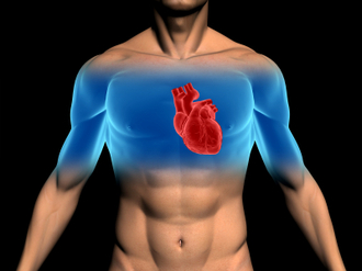
    Pericardial effusion occurs when there is an abnormal amount of fluid around the heart.  The heart is normally surrounded by a thin membranous sac called the pericardium.  The space between the pericardium and the muscle that is the heart is referred to as the perciardial space.  Normal levels of pericardial fluid within the pericardial space are from 15 to 50 mL, or about 1-3 tablespoons.

    iStock_000003834803XSmall.jpg

    An effusion, therefore, represents  an abnormal accumulation of fluid in the pericardial space.   Because of the limited amount of space in the pericardial cavity, fluid accumulation will lead to increased intrapericardial pressure and this can negatively affect heart function.  Cardiac tamponade occurrs when there is a  large enough pericardial effusion causing enough pressure to adversely affect heart function.  This is an emergent life threatening condition.

    Pericardial effusion symptoms may include difficulty breathing (dyspnea), shortness of breath when lying down (orthopnea), chest pain, cough, dizziness, low grade fever, rapid heart rate (tachycardia), and a feeling of anxiety

    Pericardial effusion may be caused by:

    -a disturbed equilibrium between the production and re-absorption of pericardial fluid,
    -a structural abnormality that allows fluid to enter the pericardial cavity
    -inflammation of the pericardium (pericarditis)
    -bacterial or viral infections
    -injury to the heart from a medical procedure
    -cancer
    -heart attack
    -autoimmune disorders

    Unfortunately, pericardial effusion and cardiac tamponade may result from improper placement of a central venous catheter during a medical procedure involving an infant.  This may arise when there is an inadvertent perforation into the pericardial space by the CVC and fluids are artificially infused into the space thereby causing the tamponade. Upon recognition of this situation, emergent removal of this fluid via a needle inserted through the chest wall and into the pericardial space (pericardiocentesis) can improve the infant’s chance of survival. It is suggested that routine radiography be performed to readily identify the CVC tip in all cases when these lines are placed into babies.    Increased awareness of this complication may decrease the mortality associated with CVC related pericardial effusions.

    Treatment depends on the underlying cause and the severity of the heart impairment. Pericardial effusion due to a viral infection sometimes goes away within a few weeks without treatment.  Some pericardial effusions remain small and never need treatment.  If the pericardial effusion is due to an autoimmune condition treatment with anti-inflammatory medications may help. If the effusion is compromising heart function and causing cardiac tamponade, it will need to be drained, most commonlyby a pericardiocentesis.  In some cases, surgical drainage may be required by cutting through the pericardium creating what is referred to as a pericardial window.


  2. Small Bowel Obstruction and Medical Malpractice

    Posted by Dr. Larry Leichter on April 15, 2018

    The small bowel is a long coiled hollow tube, called a tract, that is approximately twenty-five feet long. It includes the duodenum, jejunum and ileum.  A small bowel obstruction, also known as a small intestinal obstruction, is a mechanical or functional (paralytic) blockage of the intestinal tract, which prevents the normal transit of digestive products. It can occur at any level throughout the jejunum and ileum, and is considered a medical emergency when it occurs. The condition is often treated conservatively for the first several days; however, the patient must be monitored very closely for signs of clinical deterioration that can become life threatening.

    iStock_000017493981XSmall.jpg


    Mechanical obstruction is due to a mechanical barrier, such as an adhesive band from prior surgery, which creates a road block to the bowel.  On the other hand, functional obstruction is caused by an event that interferes with the nervous innervation of the bowel, such as electrolyte imbalances and metabolic disturbances. Functional bowel obstruction can be caused by a multitude of conditions whereas mechanical SBO is generally credited to a luminal, mural, or extra-mural mechanical barrier. A clinical syndrome exists called small intestinal pseudo-obstruction, which is characterized by manifestations of mechanical bowel obstruction in the absence of an obstructive lesion.

    The symptoms of a mechanical small bowel obstruction include abdominal fullness and/or excessive gas, abdominal distention, pains and cramps in the stomach area (specifically the mid abdomen), vomiting, constipation (inability to pass gas or stool), diarrhea, and bad breath. Acute functional small bowel dilatation is referred to as adynamic or paralytic ileus. The symptoms of paralytic obstruction, in reference to the ileus, are abdominal fullness and/or excessive gas, abdominal distention, and vomiting after eating.  The pain less closely resembles the colicky type seen in mechanical obstruction, but may be just as severe.

    The diagnosis is determined by listening to the abdomen with a stethoscope. High-pitched, tinny and clanking sounds can be heard at the onset of mechanical obstruction.  If the blockage persists for too long or the bowel is significantly damaged, due to the stretching of the blood vessels supplying it thereby decreasing blood flow, bowel sounds will decrease and eventually become silent.  The hallmark of paralytic ileus is decreased or absent bowel sounds, which can create confusion in relation to the issue of etiology if this occurs.Diagnostic tests that demonstrate obstruction include plain radiographic film of the abdomen (usually in the flat and upright position), CT scan, barium enema and upper GI series with small bowel follow through.

    Treatment depends on the cause of the obstruction. In some cases, drastic measures are necessary to save a person’s life, while in others a strategy of watchful waiting is more appropriate. In general, more serious cases that require immediate treatment can be identified based on a patient’s vital signs and physical exam. If the person is very sick and appears to be on the brink of a serious event, surgery may be required to ensure the patient’s life.

    To determine if there is any deterioration consistent with lack of blood flow, which leads to bowel ischemia, gangrene, perforation, septic shock, and death, it is imperative that the following steps be taken. The bowel must be decompressed with a long indwelling tube, all oral feeding must be stopped and IV therapy must be initiated with continuous monitoring and observation. Generally speaking, there is no reason anyone presenting to the emergency room with a small bowel obstruction should die in the hospital unless there are extenuating circumstances.