Medical Malpractice Attorneys
  • Toll Free 1-800-529-9120
  1. Brain Injury and Medical Malpractice

    Posted by Dr. Jack Sacks on May 24, 2018.

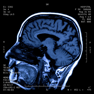
    The prevalence of brain injury in the United States is alarming as it is the second leading cause of disability in the country. Often referred to as the silent epidemic, approximately 3.17- 5.3 million Americans suffer from traumatic brain injuries, another 4.7 million have brain injuries from strokes, and another 500,000 have cerebral palsy (brain injury due to an event of oxygen deprivation). Causes of brain injury that may give rise to a medical malpractice lawsuit are further described below:

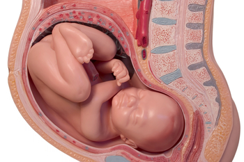
    iStock_000013877415XSmall-1-thumb-285x177-14594Brain Injury from Birth: a medical malpractice lawsuit may arise may when a child’s brain is negligently deprived of oxygen during pregnancy, labor and delivery. This may result in the child later developing cerebral palsy, mental retardation, seizures, blindness, deafness, and learning disabilities. Oxygen deprivation that injures a baby’s brain may arise from any of the following events:

    *Compression of the umbilical cord during delivery
    *Maternal Infection present during the pregnancy or delivery
    *Placental abruption or uterine rupture prior to birth
    *Maternal high blood pressure during the pregnancy (preclampsia)
    *Breeched vaginal position of the baby (feet first rather than head first)
    *Improper administration or doasage of epidural or labor inducing drugs during the delivery
    *Failure to timely perform an emergency c-section
    *Fetal macrosomia (oversized baby) unable to navigate the birth canal 

    Brain Injury in Adults and Children: a medical malpractice lawsuit may arise as a result of errors in diagnosis and treatment of a serious medical condition. A few of the causes of brain injury in children and adults that may involve medical malpractice include:

    *Medication errors
    *Anesthesia errors
    *Surgical errors
    *Radiology errors 
    *Emergency room errors
    *Delay in diagnosis/treatment of heart attack or cardiac arrest
    *Delay in diagnosis/treatment of a stroke, aneurysm, or blood clot
    *Delay in diagnosis/treatment of meningitis or encephalitis
    *Delay in diagnosis/treatment of a spreading infection or abscess
    *Delay in diagnosis/treatment of internal bleeding
    *Delay in diagnosis/treatment of hydrocephalus 
    *Delay in diagnosis/treatment of diabetes/diabetic coma/insulin shock

    In addition to the above mentioned causes of brain injury, many other errors involving patient treatment and care may give rise to a lawsuit.  One of the most common causes occurs when a hospital patient or nursing home patient falls (due to inadequate protective measures) and the patient suffers a traumatic brain injury.  In fact, falls are the leading cause of traumatic brain injury in our country surpassing even motor vehicle accidents. For a detailed guide to the incidence, prevalence, and epidemiology of brain injury, seeEssential Brain Injury Guide prepared under the auspices of the Brain Injury Association of America.     


  2. Heart Defects In Newborns

    Posted by Dr. Jack Sacks on May 22, 2018

    Heart defects in newborns affect approximately 8 out of every 1000 births.  Causes can include genetic factors, such as those found in Down’s syndrome, or factors due to the developing baby’s environment. Certain medications, such as Paxil (Paroxetine), Zoloft (Sertraline), Wellbutrin (Bupropion), or Prozac (Fluoxetine) may be associated with cardiac birth defects in the babies of mothers who took these medications when pregnant. While some heart defects may only slightly affect a child’s quality of life, others can be very serious and may require surgical correction. Although causes of many heart defects are not known, improvements in prenatal diagnosis has led to better management and outcomes for those babies affected by these conditions.

    The most common way to diagnose a cardiac defect prenatally is by sonogram. Many obstetricians order a screening sonogram for their patients during the 16th to 20th week of pregnancy. Part of the standard practice for the sonographer is to identify the four chambers of the baby’s developing heart.

    If the four chambers are not seen, or appear abnormal, a more specialized test, called a fetal echocardiogram may be ordered. This test involves a more detailed sonogram examination of the developing baby’s heart. Abnormalities can be further evaluated by this test method.

    There are many different cardiac anomalies that affect newborns. Only three (3) will be discussed here:

    Ventricular Septal Defect (VSD)- refers to a hole between the ventricles, the two lower chambersof the heart. The mixing of the oxygenated and deoxygenated blood through the defect can cause symptoms. Sometimes, a small VSD may close up on its own; sometimes surgery is required.

    Atrial Septal Defect (ASD)- involves a hole in the wall separating the atria, the upper chambers of the heart.

    Pulmonary Stenosis-occurs when the pulmonary artery that leads from the right ventricle to the lungs is narrowed. This can obstruct this crucial bloodflow out of the heart and can cause the right ventricle to become over developed and enlarged in trying to overcome the obstructive narrowing.

    If proper prenatal testing was not done, or if testing was not read or interpreted properly, medical malpractice may have occurred.  Allegations of medical malpractice may include a negligent delay in diagnosis and/or inadequate surgical treatment and care of the newborn’s heart defect.   Lawsuits may also result if a baby was injured by medications used by the mother. Cardiac defects can cause serious health problems in the child and may also involve major surgery for correction. In addition, complications can occur due to the defect, especially if undiagnosed. It is very important that women at risk for having a baby with a heart defect have appropriate screening tests done. Risk factors for congenital heart defects may include prenatal exposure to the medications listed above. Also, factors such as a sibling or family history of congenital heart defects may warrant increased surveillance and testing during the pregnancy.


  3. Advanced Diagnostic Imaging for Acquired Brain Injury

    Posted by Dr. Jack Sacks, Esq.on March 22, 2016

    One of the first steps in evaluating brain injury is diagnostic imaging. Imaging refers to various methods of viewing the structures and processes residing in the brain. Some of the more familiar modalities are CT (or CAT) scans, which use X-rays to evaluate intracranial structures. MRI, Magnetic Resonance Imaging, uses magnetic fields to illustrate the brain. However, in cases of traumatic brain injury (TBI), more advanced methods may be needed for proper diagnosis.

    iStock_000000113655XSmall.jpg

    An MRI machine can use special software to perform a brain scan called Diffusion Tensor Imaging (DTI). This scan detects the diffusion of water across brain cells and highlights certain areas that may be associated with injury. These injuries may not be apparent on conventional MRI’s.

    PET scan (Positron Emission Tomography) measures uptake and metabolism of glucose from a small radioactive “tagged” sample injected into the patient.  The scanner monitors this sample as the brain utilizes it. The metabolic uptake and usage may differ in patients who have suffered a brain injury. This helps define the extent and type of injury.

    In SPECT (Single Photon Emission Computed Tomography), the tagged sample is not absorbed or utilized in the brain cells. Instead, it remains in the blood stream and demonstrates the blood movement or perfusion through the brain. Areas of brain injury or damage may not have normal blood flow so the SPECT scan helps define these areas.

    Brain injuries can result from medical malpractice. A baby may suffer birth trauma during labor and delivery. A child may have a concussion or sports injury misdiagnosed or improperly evaluated by a physician. An individual injured in a car accident may not have the indicated testing done by the emergency room. Although scientific progress in imaging studies has improved the ability to diagnose and evaluate brain injuries, these tests need to be utilized in the appropriate situations so patient results and outcomes can improve.


  4. Shoulder Dystocia May Cause Erb’s Palsy or Brachial Plexus Injury

    Posted by Dr. Jack Sacks, Esq.on March 15, 2016

    In the typical birth of an infant, the baby arrives head first, followed by the shoulders and the rest of the body. However, in certain circumstances, the head delivers but the shoulders cannot fit through easily. This is called “shoulder dystocia” and can be an obstetrical emergency. The umblilical cord may be compressed inside the birth canal which can have grave consequences for the baby if this situation is not quickly remedied.

    iStock_000014328103XSmall.jpg

    One risk factor for shoulder dystocia is macrosomia, or large size of the baby. There are several well recognized risk factors for this. Maternal obesity may play a role. Moms with diabetes also have a higher incidence of having large babies.  In order to help minimize the incidence of these conditions special attention should be paid to mothers with these risk factors during prenatal care. Diabetes screening and management should be carefully monitored. Sonograms and fetal measurements can help with estimates of the baby’s weight. If a shoulder dystocia develops during labor, there are special steps that can be taken at that critical time to try to deliver the baby in the least traumatic fashion.

    Sometimes nerve injuries can occur in infants who have suffered a shoulder dystocia, one such unfortunate condition is called “Erb’s palsy.” In this condition, the baby has a paralysis of the arm and/or hand due to nerve damage to the brachial plexus of nerves. While some infants may improve or recover without additional treatment, many may need special procedures and even surgery to recover some function.

    Medical malpractice can arise in situations where the mother has not been properly screened for gestational diabetes, or the diabetes was not properly managed. There are situations where test results may have indicated an excessively large baby but a C-Section was not done or offered. Also, the proper precautions or techniques may not have been used during labor with a shoulder dystocia and nerve damage or Erb’s palsy may have resulted.


  5. Brain Injuries and Birth

    Posted by Dr. Jack Sacks, Esq.on March 01, 2016

    The brain is responsible for higher motor and sensory functions. It requires a constant source of oxygen in order to continue its vital functions. When the flow of oxygen is interrupted, the consequences can be devastating.  Severe injury can occur when the brain lacks the oxygen needed to continue functioning. A total lack of oxygen can be referred to as “anoxic” injury whereas a partial lack of flow can be referred to as “hypoxic” injury.

    Brain injury can occur in an unborn baby if the flow of oxygen from the mother’s circulation to the baby’s is interrupted. This can happen from a number of causes. For example, the placenta can become partially detached from the wall of the uterus (placental abruption), which can interrupt the flow of oxygen to the baby. This complication can arise in pregnant patients with untreated or inadequately treated high blood pressure. Close monitoring of mother and baby, along with timely delivery and/or C-Section when needed can prevent these complications.

    iStock_000000195410Large.jpg

    Excessive contractions of the uterus (sometimes called “uterine hyperstimulation” or “tetanic contractions”) from labor, or from medicines used to accelerate labor, like pitocin, can cause problems with the baby’s oxygen supply. The rapid, powerful contractions of the uterus can prevent maternal oxygen from reaching the baby. If the flow of oxygen to the baby is interrupted, hypoxic or anoxic brain injury can occur. The consequences of this can be severe and may include seizures, brain damage, developmental delay, cerebral palsy, and other problems with motor or cognitive functions. Use of medications like pitocin must be closely monitored by dosage and effect on the mother and baby. If contractions are too strong or too frequent, the dosage may need to be decreased, the medicine may need to be stopped entirely, or special medication to reverse the effects may be given.

    Brain injuries may occur in infants, children, and adults. Stroke, cardiac arrest, or choking can all interrupt the flow of oxygen to the brain. Patients who are undergoing surgery, or are in an intensive care unit in a hospital often have their oxygen levels monitored to be sure they are getting a sufficient supply. If they are not properly monitored or complications occur, hypoxic or anoxic brain injuries may result.

    Medical malpractice that results in brain injury is devastating for the victim as well as families and loved ones. The extent of disability from hypoxic or anoxic brain injury varies greatly, but many people require extensive care and rehabilitation to regain function and improve their quality of life.


  6. Persistent Pulmonary Hypertension of the Newborn

    Posted by Dr. Jack Sacks, Esq.on September 09, 2015

    Persistent pulmonary hypertension of the newborn (PPHN) can occur in babies when the circulatory system does not adapt to conditions outside the mother’s uterus. Before birth, the baby’s oxygen needs are met through the placenta in the mother’s uterus. After birth, the baby’s lungs take over this function. After being born, the baby inflates its lungs with its first breaths. Then, a series of changes take place to decrease resistance to blood flowing through the newborn’s lungs. This allows the lungs to take over getting oxygen for the baby’s vital functions.

    Sometimes, the changes that allow blood to preferentially flow from the baby’s heart to the lungs do not proceed normally, or at all. This can occur in approximately 2 out of 1000 births.

    Some PPHN cases are due to problems in the baby’s lung tissue. For example, if baby has damaged lungs from inhaling meconium, or pulmonary damage from prematurity, this can contribute to pulmonary hypertension. Development problems or defects in lung and related blood vessels can also lead to PPHN. However, PPHN can develop even when the lung tissue and blood vessels are normal.

    Some cases of persistent pulmonary hypertension of the newborn have been linked to medications used by pregnant women. Antidepressants such as Zoloft (sertraline), Paxil (paroxetine), Prozac (fluoxetine), Lexapro (escitalopram) and Celexa (citalopram) may be associated with PPHN. The FDA issued a public health advisory in 2006 about the use of these medications in pregnant women.

    It is crucial for pregnant women to review their medical and medication histories with their doctors. Certain medications may need to be adjusted, stopped, or continued according to the patient’s needs and risks to the unborn baby. If potentially damaging medications were used and a birth defect such as PPHN occurred, the victims may have legal rights that need to be protected.


  7. Congenital Diaphragmatic Hernia in Newborns

    Posted by Dr. Jack Sacks, Esq.on February 27, 2015

    Hernia is when an organ or tissue protrudes through a weak area in the surrounding muscle or connective tissue. There are different types of hernias that generally vary by location. Congenital diaphragmatic hernia (CDH) is a developmental defect of the diaphragm that permits the abdominal viscera to herniate into the chest. The amount of herniated contents may be small or large and it generally contains parts of the intestine, spleen, or liver. CDH occurs during a critical period of lung development when bronchial and pulmonary artery branching occurs so lung compression by herniated bowel can lead to pulmonary hypoplasia, which is underdevelopment of the lungs. CDH occurs in 1 out of 2200 births and in 50% of CDH cases, there are other associated anomalies such as chromosomal abnormalities, congenital heart disease, and neural tube defects.

    Within the first few hours of life, infants with CDH may present with mild or severe respiratory distress that may be incompatible with life. Symptoms include bluish skin due to lack of oxygen, rapid breathing, and fast heart rate. On physical exam, patients may have a barrel-shaped chest, scaphoid appearing abdomen due to loss of abdominal contents into the chest, and absence of breath sounds on the affected side. In the majority of patients with CDH, herniation occurs on the left; therefore, the heartbeat is displaced to the right due to a shift in the mediastinum. Right sided diaphragmatic hernias occur in 11% of cases and bilateral herniation only in 2%.

    The level of respiratory distress depends on the severity of lung hypoplasia. In the prenatal period, lung hypoplasia can be determined using ultrasound to evaluate herniated contents and to measure the lung area to head circumference ratio. Postpartum, there is no specific test to quantify the amount of hypoplasia. The diagnosis of CDH is generally made prenatally by ultrasound; however, in those where CDH is not diagnosed in utero, it should be suspected in any full term infant presenting with respiratory distress and the diagnosis is made by chest x-ray showing herniation of abdominal contents into the hemithorax.

    For neonates diagnosed by prenatal ultrasound, the following steps for monitoring and intervention are recommended: twice weekly nonstress testing or biophysical profile testing at 33-34 weeks in addition to ultrasound examinations at 28, 30, 32, and 34-35 weeks to assess fetal growth and amniotic fluid volume. If the fetus experiences growth restriction or oligohydramnios, which is a deficiency of amniotic fluid, the baby should deliver early and betamethasone (a steroid medication that assists in preterm fetal lung development) should be given prior to delivery if the fetus is less than 34 weeks.  The best mode and gestational age for delivery of a fetus with CDH is undetermined; however, the suggested time for planned induction of labor is between 38-39 weeks.

    In the delivery room, infants with problematic CDH should be immediately intubated and ventilated with low peak pressure to minimize lung injury. A nasogastric tube on continuous suction is placed in the stomach for decompression of the abdominal contents, which can help expand available lung tissue. In addition, the infant should have an umbilicial artery line for monitoring of blood gases and blood pressure and possibly an umbilical vein catheter for administration of fluids and medications. Blood pressure support should be given and an echo should be done to determine cardiac abnormalities in addition to the extent of pulmonary hypertension and shunting.

    Extracorporeal membrane oxygenation (ECMO) has been used as part of  the treatment in some hospitals. ECMO operates as a heart-lung bypass system; thus, it does the job the heart and lungs would be doing. ECMO can be used temporarily while the infant’s condition stabilizes and improves.  Once the infant is stable, he or she can undergo surgical repair of the diaphragmatic hernia, where the stomach, intestines, and other abdominal organs are returned to the abdominal cavity. The hole in the diaphragm is also repaired. If the diaphragm is absent, an artificial diaphragm will be constructed and placed. Following the operation, the infant will require breathing support due to underdevelopment of the lungs. Once the infant is taken off ventilation (breathing machine), he or she may need oxygen and medications to assist with breathing for weeks to years. Thus, CDH requires long term follow up to monitor the infant’s condition to ensure no future complications such as lung infections or other associated congenital problems. The prognosis is generally good for infants with CDH and survival is greater than 80%.


  8. Child Abuse and Neglect

    Posted by Dr. Jack Sacks, Esq.on January 02, 2013

    Child neglect is the most prevalent form of child maltreatment in the US. According to the National Child Abuse and Neglect Data System (NCANDS), approximately 899,000 children in the US who were victims of abuse and neglect in 2005, 62.8% suffered from neglect alone. Child neglect is a failure by parents or caregivers to provide for a child’s basic physical, educational, and emotional needs. It is difficult to define because it varies with the age and development of the child; therefore, it is problematic to determine the extent of the problem. Risk factors for abuse include: preexisting mental or physical illness, poverty, familial stress, substance abuse by parents or caregives. Physicians, nurses, day care personnel, relatives, and neighbors are generally the ones to suspect and report neglect; however, they may be the culprits too. Often times, signs of child neglect may include inadequate medical or custodial care.

    Physical neglect encompasses the majority of neglect cases and it involves the parent/caretaker not granting the child basic necessities such as food, clothing, medical care, and shelter. By depriving the child of these essential things, his or her physical health, well being, psychological growth and development is jeopardized. Failure to thrive, malnutrition, serious illness, physical injury due to lack of supervision, and low self-esteem are a some of the byproducts of physical neglect. As an unfortunate example, infants are sometimes mistakenly left behind in cars or vans by their caregivers when the outside temperature is very hot, resulting in heat stroke or death to these infants.

    Regarding child abuse, medical neglect is the failure to deliver necessary healthcare for a child thereby endangering the child’s well being. Situations involving medical neglect include when a parent refuses medical care for a child in an emergency or when a parents does not follow recommendations for a child with a treatable chronic disease or disability. Medical neglect often correlates with poverty; however, there is a difference between an inability to provide the appropriate care due to finances or cultural norms and being purposefully neglectful.

    If one suspects child neglect, the first step is to report the incident to local child protective services agency in the county or state. The majority of states have a child abuse hotline or you can use the Childhelp National Child Abuse Hotline (1-800-4-A-CHILD). Professionals involved with children have a duty to report reasonable suspicion of abuse and neglect. Reasonable suspicion is based on objective evidence which includes firsthand observation or statements made by a parent or child. Symptoms of negelect include: difficulties in school, eating disorders, low self esteem, depression, anxiety, signs of physical injury, rebellious behavior, sleep disorders, and other vague physical complaints.


  9. Pituitary Adenomas and Medical Malpractice

    Posted by Dr. Jack Sacks, Esq.on October 25, 2011

    A pituitary adenoma is generally a benign, slow growing tumor that occurs in the pituitary gland. The pituitary gland is a small, bean shaped structure that lies at the base of the brain. It has a central role in the regulation of hormones that affect the body such as Adrenocorticotropic hormone (ACTH), Growth hormone (GH), Prolactin, and Thyroid-stimulating hormone (TSH).

    Approximately 1 in 1,000 individuals have pituitary adenomas. They are generally not cancerous but may invade nearby structures.  They are classified based on size. A microadenoma is less than 1 cm in diameter whereas a macroadenoma is larger than 1 cm in size.

    iStock_000017548218XSmall.jpg

    Based on whether the pituitary adenoma is a hormone-producing or hormone-inactive tumor, the patient will present with different symptoms. Hormone-producing tumors will make excessive amounts of an active hormone so symptoms present as a hormonal imbalance. The three most common hormone-producing adenomas are Prolactinomas, Growth hormone-secreting pituitary adenoma, and ACTH-secreting pituitary adenoma.

    If the patient has a large hormone-inactive or hormone-producing tumor, it may compress surrounding brain structures due to its size.  Large pituitary tumors may compress the pituitary gland contributing to pituitary failure, which can lead to sexual dysfunction, inadequate body cortisol levels, and hypothyroidism. Other possible presentations relating to compression of brain structures include visual loss, headache, the “stalk effect”, and pituitary apoplexy.  The “stalk effect” is due to the compression of the pituitary stalk, the structure connecting the brain to the pituitary gland, which leads to a mild elevation in the hormone prolactin. Higher levels of prolactin in females can contribute to irregular menstrual cycles. Pituitary apoplexy presents with an abrupt headache and visual loss. It can occur under two different situations. The pituitary adenoma can bleed internally causing a sudden increase in size or the tumor can outgrow its blood supply and the dead tissue will swell.

    Pituitary adenomas can be diagnosed based on endocrine function testing, imaging, and visual field testing. Often times, visual problems may be the only symptom present.  Optometrists and ophthalmologists should seriously consider the possibility of a pituitary tumor when the patient presents with an unexplained loss of visual field (especially peripheral vision), double vision, or blurred vision as the growth of a pituitary tumor can cause compression on the visual pathway (retina, optic nerve, etc). A delay in diagnosis of a pituitary adenoma may result in permanent loss of vision due to the damage caused by this compression. Endocrine function testing evaluates cortisol, follicle-stimulating hormone, lutenizing hormone, insulin growth factor-1, prolactin, testosterone/estradiol, and thyroid hormone levels. The preferred method of imaging is MRI, which screens for adenomas larger than 4 mm.

    Treatment for pituitary adenomas depends on the presence of hormone production, size of the tumor, invasion of the tumor into surrounding structures, and the age and health of the patient. Drug therapy is used to treat hormone-producing tumors. For example, bromocriptine and cabergoline are used to treat tumors secreting prolactin because these medications decrease prolactin levels and tumor size. Pituitary adenomas that require surgery are usually minimally invasive techniques, where the tumor is removed through the nose.  On the other hand, radiation therapy involves high doses of radiation being delivered to the tumor. It is a treatment utilized for pituitary adenomas that cannot be controlled by drug therapy or surgical intervention. Patients have the best outlook when the entire tumor can be removed.


  10. Premature Birth May Result in Injury to the Newborn

    Posted by Dr. Jack Sacks, Esq.on August 09, 2011

    Premature birth can often result in complications for the newborn. Preterm or premature birth refers to babies born before 37 weeks of a normal 40 week long pregnancy.

    Some complications that can affect babies born prematurely are lung complications from lack of pulmonary maturity or development. Eye problems can result in vision difficulties or blindness. Premature babies may be more prone to infections and intestinal problems. They also are at increased risk for bleeding in the brain which can have devastating effects. Conditions ranging from learning disabilities to cerebral palsy and seizures are more common in babies born prematurely.

    Thus it is extremely important that premature labor be diagnosed in a timely fashion by the patient’s health care provider. Steps can be taken to treat the underlying cause of the preterm labor. For example, if a maternal infection is present that is related to the preterm contractions , the infection needs to be diagnosed and treated. Certain conditions, such as twins, increase the risk of premature labor. Also, some anatomic abnormalities of the cervix or uterus can elevate the risk as well.

    Although it may not be possible to prevent premature birth, certain steps can be taken to prolong the time until delivery. This may give time for medications to be administered that may significantly improve the newborns outcome by enhancing lung development or decreasing the risk of intestinal problems. Infections that are properly diagnosed and treated may likewise improve the premature baby’s situation.

    Medical malpractice may occur when a pregnant patient’s symptoms, such as premature contractions, bleeding, or rupture of membranes are not properly evaluated and managed. If needed interventions such as medication to improve lung function, or antibiotics, are not given in cases where they are indicated, malpractice may have occurred. An evaluation by attorneys with experience and knowledge of these complex medical and legal issues can help clarify issues for victims of medical malpractice.