Medical Malpractice Attorneys
  • Toll Free 1-800-529-9120
  1. Deep Venous Thrombosis and Medical Malpractice

    Posted by Dr. Jack Sacks, Esq.on March 28, 2016

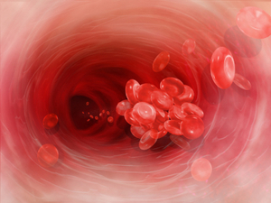
    Deep venous thrombosis is the development of a blood clot in the large, deep veins of the lower leg and thigh. Thrombi can cause tissue injury due to vascular occlusion or distal embolization. However, venous obstruction can be offset by collateral blood vessels. Thrombi can also cause local pain and edema due to the blockage of blood flow. If the clot breaks off and travels through the blood, it is referred to as an embolism. An embolism can become trapped in the brain, lungs, or heart leading to major injury. Pulmonary embolus (PE) is a common complication and life threatening if not treated quickly with anticoagulants. PE presents with shortness of breath, chest pain, and cough with blood in sputum.

    iStock_000012053156XSmall.jpgDVT can occur with stasis or in hypercoaguable states. It is commonly seen following trauma, surgery, or burns, which contribute to decreased physical activity, damage to vessels, and release of procoagulant substances from tissues. Reduced physical activity causes a decline in the milking action of lower leg muscles and slows venous return. Risk factors for DVT include advanced age, bed rest, immobilization, smoking, birth control pills, family history of blood clots, fractures in the pelvis or legs, giving birth within the last 6 months, heart failure, and obesity.  To prevent DVT, patients should move their legs during long flights or when they are immobile for long periods of time.

    Although many DVTs are asymptomatic, they can recur. Some individuals suffer from post-phlebitic syndrome, which involves chronic pain and swelling in the leg. The major symptoms of DVT include changes in a patient’s leg such as redness, increased temperature, pain, and tenderness. Diagnosis is based on the physical exam, which should demonstrate a red, swollen leg. Diagnostic tests include a D-dimer blood test along with other blood tests to check for hypercoagulability such as activated protein C resistance, anti-thrombin III levels, antiphospholipid antibodies, and genetic testing for mutations with a predisposition towards blood clots. Imaging studies of the legs include Doppler ultrasound, plethysmography, and radiography.

    The primary treatment for DVT is anti-coagulants, also known as blood thinners. They prevent the formation of new clots and the growth of old clots. However, they cannot dissolve existing clots. Patients are more likely to bleed on these medications. Heparin is an IV administered anticoagulant given in a hospital setting.  Warfarin (Coumadin) is an oral anticoagulant that takes several days to work; thus, Heparin cannot be stopped until Warfarin is functioning at an effective dose for a minimum of two days. Many patients wear pressure stockings on their legs to improve blood flow and decrease their risk of DVT.  When medications are ineffective, patients may need to undergo surgery. A filter can be placed in the body’s largest vein to prevent thrombi from migrating to the lungs. Also, surgery may be necessary to remove large thrombi.


  2. Pulmonary Embolism and Medical Malpractice

    Posted by Dr. Jack Sacks, Esq.on March 20, 2016

    Pulmonary embolism (PE) is a life threatening condition that affects over 600,000 people per year in the United States. Through the use of modern diagnostic tools such as multislice spiral CT and advancements in treatment, the mortality rate of PE has been reduced in recent years.

    iStock_000000529518XSmall.jpg

    PE involves a blockage of an artery in the lungs from a clot that has traveled through the bloodstream from another part of the body. Usually the clot begins in the deep veins of the legs where it is referred to as deep vein thrombosis or DVT. The risk of PE increases during periods of immobilization such as long plane flights, surgery, pregnancy, and with conditions such as cancer and obesity. Additionally, estrogen based hormonal contraception (birth control) may increase the risk of PE.

    Signs and symptoms of PE may include chest pain, shortness of breath, abnormal EKG (such as inverted T-waves), rapid heart rate, and increased respirations. PE may be preceded by leg pain due to DVT. A low grade fever may be present as well as a cough or hemoptysis (coughing up blood). If PE is suspected, a D-dimer blood test should be performed which measures the level of specific clotting related protein fragments in the blood.  The D-dimer test can help rule out PE if the blood test comes back within normal limits. However, If the D-dimer blood test comes back elevated for suspected PE, then radiographic imaging is indicated.

    When available for imaging, the multi-slice spiral CT is favorable because it is non-invasive and is highly predictive of PE. If the patient has leg pain or other signs of DVT, then a ultrasound (doppler) of the veins in the legs may be performed as there is a high correlation between DVT and PE. But a negative leg doppler does not rule out PE. Other studies to help diagnose PE include include ventilation perfusion scanning and CTPA (CT Pulmonary Angiography-with contrast).

    Treatment for PE depends on the severity or size of the blockage. For severe cases that are emergent, thrombolysis may be the preferred treatment- this involves the administration of clot busting medication such as tPA. In specific situations of severe emergent PE, surgery (embolectomy) may be an option to remove the clot. In most cases of PE, anticoagulants such as heparin and warfarin are started early as possible and used to treat the condition. Heparin or LMWH (low molecular weight heparin) are typically given initially followed by warfarin.  When indicated, an IVC filter may be placed in the patient to help prevent further clots from forming in the arteries of the lungs.


  3. Leg Amputation and Medical Malpractice

    Posted by Dr. Jack Sacks, Esq.on January 02, 2016

    Amputation is the surgical removal of all or part of an extremity. The most common amputation surgery is above or below the knee.  The indications for leg amputation include severe trauma, significant tumor in the bone or muscle, lack of blood circulation due to peripheral arterial disease, worsening or uncontrollable infection, failed management of acute compartment syndrome, failed management of Charcot’s degenerative osteoarthropathy, or debilitating extremity paralysis from infection or pressure-related complications. Failute to timely diagnose and treat infection, tumor, pressure sores, vascular disease, compartment syndrome, and Charcot’s all may result in the unneccessary amputation of a leg and give rise to allegations of medical malpractice against negligent health care practitioners.

    iStock_000026640818XSmall.jpg

    The amputation procedure varies depending on the extremity undergoing the operation. To determine the operation site and the amount of tissue to remove, the surgeon relies on the following factors: the patient’s pulse, skin temperature, areas of reddened skin, and sensitivity to touch in the affected extremity. The presence of a palpable pulse proximal to the level of amputation is a positive predictor for successful healing; however, the absence of a pulse does not necessarily reflect future wound healing failure. The level of the amputation is based on the extent of the damaged tissue, the healing potential of the area, and the rehabilitation potential of the patient. In addition to a thorough clinical examination, objective tests such as ankle pressures, toe pressures, transcutaneous oxygen measurements, and skin perfusion pressures are useful.

    The preoperative evaluation and preparation involves medical risk assessment, nutrition assessment, prosthetic and rehab consultation, and possibly a psychological consultation. The diseased tissue is removed along with any crushed bone and the maximal amount of healthy tissue is left behind. The blood vessels and nerves in the surrounding area are sealed off. Following the amputation, the site can be left open due the possibility of further amputation or covered with skin flaps and closed. The remaining muscles in the area are shaped so the end of the limb can be fitted for prosthesis, also known as an artificial limb.

    Thromboprophylaxis is recommended for all patients undergoing major lower extremity amputation because patients are at high risk for thromboembolism, the blocking of a blood vessel by a particle that has separated from a blood clot at the formation site. Antibiotic prophylaxis is typically recommended within one hour of skin incision for lower extremity amputation due to high risk for surgical site infection.

    Generally, the patient undergoes physical rehabilitation soon after surgery and practice with the prosthesis can begin 10-14 days after surgery. The patient’s postoperative outcome is dependent upon preoperative functional status, comorbidities, and the level of amputation. Wound healing must be monitored and dressing changes performed. Patients with advanced diabetes, significant heart disease, or serious infection are at a greater risk of complications from the procedure. Possible complications include infection, joint contracture, necrosis, deep vein thrombosis, pulmonary embolism, hematoma, and wound opening. In addition, patients may experience phantom pain, a sense of pain in the amputated limb described as burning aching, or electric. Other causes of pain such as ischemia, infection, neuroma, or pressure related wounds should be excluded before determining the diagnosis as phantom pain.