Medical Malpractice Attorneys
  • Toll Free 1-800-529-9120
  1. Brain Injury and Medical Malpractice

    Posted by Dr. Jack Sacks on May 24, 2018.

    The prevalence of brain injury in the United States is alarming as it is the second leading cause of disability in the country. Often referred to as the silent epidemic, approximately 3.17- 5.3 million Americans suffer from traumatic brain injuries, another 4.7 million have brain injuries from strokes, and another 500,000 have cerebral palsy (brain injury due to an event of oxygen deprivation). Causes of brain injury that may give rise to a medical malpractice lawsuit are further described below:

    iStock_000013877415XSmall-1-thumb-285x177-14594Brain Injury from Birth: a medical malpractice lawsuit may arise may when a child’s brain is negligently deprived of oxygen during pregnancy, labor and delivery. This may result in the child later developing cerebral palsy, mental retardation, seizures, blindness, deafness, and learning disabilities. Oxygen deprivation that injures a baby’s brain may arise from any of the following events:

    *Compression of the umbilical cord during delivery
    *Maternal Infection present during the pregnancy or delivery
    *Placental abruption or uterine rupture prior to birth
    *Maternal high blood pressure during the pregnancy (preclampsia)
    *Breeched vaginal position of the baby (feet first rather than head first)
    *Improper administration or doasage of epidural or labor inducing drugs during the delivery
    *Failure to timely perform an emergency c-section
    *Fetal macrosomia (oversized baby) unable to navigate the birth canal 

    Brain Injury in Adults and Children: a medical malpractice lawsuit may arise as a result of errors in diagnosis and treatment of a serious medical condition. A few of the causes of brain injury in children and adults that may involve medical malpractice include:

    *Medication errors
    *Anesthesia errors
    *Surgical errors
    *Radiology errors 
    *Emergency room errors
    *Delay in diagnosis/treatment of heart attack or cardiac arrest
    *Delay in diagnosis/treatment of a stroke, aneurysm, or blood clot
    *Delay in diagnosis/treatment of meningitis or encephalitis
    *Delay in diagnosis/treatment of a spreading infection or abscess
    *Delay in diagnosis/treatment of internal bleeding
    *Delay in diagnosis/treatment of hydrocephalus 
    *Delay in diagnosis/treatment of diabetes/diabetic coma/insulin shock

    In addition to the above mentioned causes of brain injury, many other errors involving patient treatment and care may give rise to a lawsuit.  One of the most common causes occurs when a hospital patient or nursing home patient falls (due to inadequate protective measures) and the patient suffers a traumatic brain injury.  In fact, falls are the leading cause of traumatic brain injury in our country surpassing even motor vehicle accidents. For a detailed guide to the incidence, prevalence, and epidemiology of brain injury, seeEssential Brain Injury Guide prepared under the auspices of the Brain Injury Association of America.     


  2. Increased Fluid Around the Heart May Cause Cardiac Tamponade

    Posted by Dr. Jack Sacks on April 30, 2018

    Pericardial effusion occurs when there is an abnormal amount of fluid around the heart.  The heart is normally surrounded by a thin membranous sac called the pericardium.  The space between the pericardium and the muscle that is the heart is referred to as the perciardial space.  Normal levels of pericardial fluid within the pericardial space are from 15 to 50 mL, or about 1-3 tablespoons.

    iStock_000003834803XSmall.jpg

    An effusion, therefore, represents  an abnormal accumulation of fluid in the pericardial space.   Because of the limited amount of space in the pericardial cavity, fluid accumulation will lead to increased intrapericardial pressure and this can negatively affect heart function.  Cardiac tamponade occurrs when there is a  large enough pericardial effusion causing enough pressure to adversely affect heart function.  This is an emergent life threatening condition.

    Pericardial effusion symptoms may include difficulty breathing (dyspnea), shortness of breath when lying down (orthopnea), chest pain, cough, dizziness, low grade fever, rapid heart rate (tachycardia), and a feeling of anxiety

    Pericardial effusion may be caused by:

    -a disturbed equilibrium between the production and re-absorption of pericardial fluid,
    -a structural abnormality that allows fluid to enter the pericardial cavity
    -inflammation of the pericardium (pericarditis)
    -bacterial or viral infections
    -injury to the heart from a medical procedure
    -cancer
    -heart attack
    -autoimmune disorders

    Unfortunately, pericardial effusion and cardiac tamponade may result from improper placement of a central venous catheter during a medical procedure involving an infant.  This may arise when there is an inadvertent perforation into the pericardial space by the CVC and fluids are artificially infused into the space thereby causing the tamponade. Upon recognition of this situation, emergent removal of this fluid via a needle inserted through the chest wall and into the pericardial space (pericardiocentesis) can improve the infant’s chance of survival. It is suggested that routine radiography be performed to readily identify the CVC tip in all cases when these lines are placed into babies.    Increased awareness of this complication may decrease the mortality associated with CVC related pericardial effusions.

    Treatment depends on the underlying cause and the severity of the heart impairment. Pericardial effusion due to a viral infection sometimes goes away within a few weeks without treatment.  Some pericardial effusions remain small and never need treatment.  If the pericardial effusion is due to an autoimmune condition treatment with anti-inflammatory medications may help. If the effusion is compromising heart function and causing cardiac tamponade, it will need to be drained, most commonlyby a pericardiocentesis.  In some cases, surgical drainage may be required by cutting through the pericardium creating what is referred to as a pericardial window.


  3. Small Bowel Obstruction and Medical Malpractice

    Posted by Dr. Larry Leichter on April 15, 2018

    The small bowel is a long coiled hollow tube, called a tract, that is approximately twenty-five feet long. It includes the duodenum, jejunum and ileum.  A small bowel obstruction, also known as a small intestinal obstruction, is a mechanical or functional (paralytic) blockage of the intestinal tract, which prevents the normal transit of digestive products. It can occur at any level throughout the jejunum and ileum, and is considered a medical emergency when it occurs. The condition is often treated conservatively for the first several days; however, the patient must be monitored very closely for signs of clinical deterioration that can become life threatening.

    iStock_000017493981XSmall.jpg


    Mechanical obstruction is due to a mechanical barrier, such as an adhesive band from prior surgery, which creates a road block to the bowel.  On the other hand, functional obstruction is caused by an event that interferes with the nervous innervation of the bowel, such as electrolyte imbalances and metabolic disturbances. Functional bowel obstruction can be caused by a multitude of conditions whereas mechanical SBO is generally credited to a luminal, mural, or extra-mural mechanical barrier. A clinical syndrome exists called small intestinal pseudo-obstruction, which is characterized by manifestations of mechanical bowel obstruction in the absence of an obstructive lesion.

    The symptoms of a mechanical small bowel obstruction include abdominal fullness and/or excessive gas, abdominal distention, pains and cramps in the stomach area (specifically the mid abdomen), vomiting, constipation (inability to pass gas or stool), diarrhea, and bad breath. Acute functional small bowel dilatation is referred to as adynamic or paralytic ileus. The symptoms of paralytic obstruction, in reference to the ileus, are abdominal fullness and/or excessive gas, abdominal distention, and vomiting after eating.  The pain less closely resembles the colicky type seen in mechanical obstruction, but may be just as severe.

    The diagnosis is determined by listening to the abdomen with a stethoscope. High-pitched, tinny and clanking sounds can be heard at the onset of mechanical obstruction.  If the blockage persists for too long or the bowel is significantly damaged, due to the stretching of the blood vessels supplying it thereby decreasing blood flow, bowel sounds will decrease and eventually become silent.  The hallmark of paralytic ileus is decreased or absent bowel sounds, which can create confusion in relation to the issue of etiology if this occurs.Diagnostic tests that demonstrate obstruction include plain radiographic film of the abdomen (usually in the flat and upright position), CT scan, barium enema and upper GI series with small bowel follow through.

    Treatment depends on the cause of the obstruction. In some cases, drastic measures are necessary to save a person’s life, while in others a strategy of watchful waiting is more appropriate. In general, more serious cases that require immediate treatment can be identified based on a patient’s vital signs and physical exam. If the person is very sick and appears to be on the brink of a serious event, surgery may be required to ensure the patient’s life.

    To determine if there is any deterioration consistent with lack of blood flow, which leads to bowel ischemia, gangrene, perforation, septic shock, and death, it is imperative that the following steps be taken. The bowel must be decompressed with a long indwelling tube, all oral feeding must be stopped and IV therapy must be initiated with continuous monitoring and observation. Generally speaking, there is no reason anyone presenting to the emergency room with a small bowel obstruction should die in the hospital unless there are extenuating circumstances.


  4. Meningitis and Medical Malpractice

    Posted by Dr. Jack Sacks, Esq.on March 28, 2016

    Meningitis is inflammation of the meninges, the membranes that cover the brain and spinal cord. It is commonly caused by infection, but other causes include chemical irritants, drug allergies, fungi, and tumors. Based on the clinical evolution of the illness and the type of inflammatory exudate present in the cerebral spinal fluid (CSF), infectious meningitis is categorized into acute pyogenic (usually bacterial), aseptic (usually viral), and chronic (usually tuberculosis, spirochetal, cryptococcal).

    iStock_000003651637XSmall.jpg

    Early diagnosis is essential for bacterial meningitis because it can result in death or brain damage if left untreated. In bacterial meningitis, a correlation exists between bacterial organism and age. The most likely organism in neonates may be Escherichia Coli or group B Streptococci. In the elderly, it may be Streptococcus Pneumonia or Listeria Monocytogenes. In young adults, it may be Neisseria Meningitides. In contrast, most viral infections are due to enteroviruses but only a small number of people who develop enteroviral infections present with meningitis. Other viral infections that can cause meningitis include mumps, herpes virus, measles, and influenza. Chronic meningitis can be caused by pathogens such as mycobacteria and spirochetes. Thus, medical attention is necessary to differentiate between bacterial, viral, and chronic meningitis.

    Risk factors include individuals over the age of 60 or below the age of 5, diabetes mellitus, renal or adrenal insufficiency, hypoparathyroidism, cystic fibrosis, immunosuppression, HIV, crowding (military recruits and college residents), recent exposure to those with meningitis, etc. The symptoms have a rapid onset and include fever, chills, mental status changes, nausea, vomiting, photophobia, severe headache, and meningismus (stiff neck). Additional symptoms include agitation, bulging fontanelles, decreased consciousness, tachypnea, poor feeding or irritability in children, and opisthotonos (unusual posture, with head and neck arched backwards).

    To confirm a diagnosis, a lumbar puncture (spinal tap) should generally be performed on anyone suspected of meningitis to sample and culture the CSF for abnormal cell counts, glucose, and protein. Other diagnostic tests include blood culture, chest x-ray, and MRI or CT scan of the head. The underlying cause of the meningitis needs to be determined to administer proper treatment and define the severity of each case. Unlike bacterial meningitis, viral meningitis usually does not involve treatment and patients generally recover within two weeks; however, in certain instances (such as with the herpes simplex virus) antiviral medications may be indicated.

    Antibiotic treatment for bacterial meningitis is dependent on the underlying bacterium. By treating the most common types, the risk of dying is reduced to below 15%. Symptoms such as brain swelling, shock, and seizures are treated with other medications and intravenous fluids. Possible complications of meningitis include brain damage, subdural effusion, hearing loss, hydrocephalus, and seizures. To prevent contraction of meningitis, the meningococcal vaccination is recommended for populations at risk.


  5. Advanced Diagnostic Imaging for Acquired Brain Injury

    Posted by Dr. Jack Sacks, Esq.on March 22, 2016

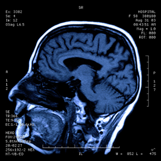
    One of the first steps in evaluating brain injury is diagnostic imaging. Imaging refers to various methods of viewing the structures and processes residing in the brain. Some of the more familiar modalities are CT (or CAT) scans, which use X-rays to evaluate intracranial structures. MRI, Magnetic Resonance Imaging, uses magnetic fields to illustrate the brain. However, in cases of traumatic brain injury (TBI), more advanced methods may be needed for proper diagnosis.

    iStock_000000113655XSmall.jpg

    An MRI machine can use special software to perform a brain scan called Diffusion Tensor Imaging (DTI). This scan detects the diffusion of water across brain cells and highlights certain areas that may be associated with injury. These injuries may not be apparent on conventional MRI’s.

    PET scan (Positron Emission Tomography) measures uptake and metabolism of glucose from a small radioactive “tagged” sample injected into the patient.  The scanner monitors this sample as the brain utilizes it. The metabolic uptake and usage may differ in patients who have suffered a brain injury. This helps define the extent and type of injury.

    In SPECT (Single Photon Emission Computed Tomography), the tagged sample is not absorbed or utilized in the brain cells. Instead, it remains in the blood stream and demonstrates the blood movement or perfusion through the brain. Areas of brain injury or damage may not have normal blood flow so the SPECT scan helps define these areas.

    Brain injuries can result from medical malpractice. A baby may suffer birth trauma during labor and delivery. A child may have a concussion or sports injury misdiagnosed or improperly evaluated by a physician. An individual injured in a car accident may not have the indicated testing done by the emergency room. Although scientific progress in imaging studies has improved the ability to diagnose and evaluate brain injuries, these tests need to be utilized in the appropriate situations so patient results and outcomes can improve.


  6. Spinal Cord Compression and Cauda Equina Syndrome

    Posted by Dr. Jack Sacks, Esq.on January 03, 2016

    There are approximately 12,000 new cases of spinal cord compression annually in the United States with the average age of victims being 39.5 years old. Causes of spinal cord compression include trauma (such as auto accidents, falls, sports injury, epidural injection), spinal abscess, tumor, hematoma or blood clot, ruptured or herniated disk, and spinal stenosis. . Early symptoms may include the start of loss of movement or feeling in the arms or legs, back pain, and the loss of bowel or bladder function or control. Spinal cord compression of sudden onset constitutes a medical emergency, as the longer the duration of symptoms before cord pressure relief is obtained by surgical intervention (laminectomy), the greater the chance of permanent injury. Diagnosis is by clinical exam and other testing such as X-ray, CT, and MRI. MRI is usually the most accurate study to detect spinal cord compression as details of both bony and soft tissue abnormalities in the spinal column may be visualized. MRI may also be the preferred study if spinal cord injury occurs during pregnancy as MRI offers reduced radiation exposure to the fetus. However, after an accident, traction devices to immobilize the spine and life support equipment may preclude the use of MRI.

    The spinal column is comprised in descending order of the cervical, thoracic, lumbar, and sacral areas. The actual spinal cord ends at the level of L-1. Below the level of L-1, the cord branches into a bundle of spinal nerve roots from L-1 through L-5 and S-1 through S-5 that resembles a horses tail referred to as the cauda equina. When the nerve roots of the cauda equina are compressed permanent neurogenic injury may occur. This may be referred to as cauda equina syndrome (CES). Causes of compression of these nerve roots include the same types of causes as listed above for spinal cord compression. Symptoms of early onset of cauda equina syndrome include numbness in the groin (saddle anesthesia), loss of bowel or bladder function or control, weakness in the legs, and absence of ankle reflexes. Early diagnosis and treatment of CES is important as the longer the duration of symptoms, the more likely permanent neurological injury will occur such as paralysis and incontinence. Cauda equina syndrome of sudden onset is a medical emergency and treatment generally involves surgical decompression of the affected nerve roots. A laminectomy may be performed to relieve pressure on the nerve root in cases where there is a herniated or ruptured disk, hematoma, abscess, or tumor.

    In spinal cord and nerve root compression cases involving sudden onset of neurological symptoms, medical malpractice may arise when there has been an unreasonable delay in diagnosis and prompt surgical intervention by the doctors or hospital. Allegations of medical malpractice may also include a failure to diagnose and treat a vertebral fracture of the neck or back that required traction or immobilization which was not performed, thereby allowing pathological movement that resulted in spinal cord compression.


  7. Leg Amputation and Medical Malpractice

    Posted by Dr. Jack Sacks, Esq.on January 02, 2016

    Amputation is the surgical removal of all or part of an extremity. The most common amputation surgery is above or below the knee.  The indications for leg amputation include severe trauma, significant tumor in the bone or muscle, lack of blood circulation due to peripheral arterial disease, worsening or uncontrollable infection, failed management of acute compartment syndrome, failed management of Charcot’s degenerative osteoarthropathy, or debilitating extremity paralysis from infection or pressure-related complications. Failute to timely diagnose and treat infection, tumor, pressure sores, vascular disease, compartment syndrome, and Charcot’s all may result in the unneccessary amputation of a leg and give rise to allegations of medical malpractice against negligent health care practitioners.

    iStock_000026640818XSmall.jpg

    The amputation procedure varies depending on the extremity undergoing the operation. To determine the operation site and the amount of tissue to remove, the surgeon relies on the following factors: the patient’s pulse, skin temperature, areas of reddened skin, and sensitivity to touch in the affected extremity. The presence of a palpable pulse proximal to the level of amputation is a positive predictor for successful healing; however, the absence of a pulse does not necessarily reflect future wound healing failure. The level of the amputation is based on the extent of the damaged tissue, the healing potential of the area, and the rehabilitation potential of the patient. In addition to a thorough clinical examination, objective tests such as ankle pressures, toe pressures, transcutaneous oxygen measurements, and skin perfusion pressures are useful.

    The preoperative evaluation and preparation involves medical risk assessment, nutrition assessment, prosthetic and rehab consultation, and possibly a psychological consultation. The diseased tissue is removed along with any crushed bone and the maximal amount of healthy tissue is left behind. The blood vessels and nerves in the surrounding area are sealed off. Following the amputation, the site can be left open due the possibility of further amputation or covered with skin flaps and closed. The remaining muscles in the area are shaped so the end of the limb can be fitted for prosthesis, also known as an artificial limb.

    Thromboprophylaxis is recommended for all patients undergoing major lower extremity amputation because patients are at high risk for thromboembolism, the blocking of a blood vessel by a particle that has separated from a blood clot at the formation site. Antibiotic prophylaxis is typically recommended within one hour of skin incision for lower extremity amputation due to high risk for surgical site infection.

    Generally, the patient undergoes physical rehabilitation soon after surgery and practice with the prosthesis can begin 10-14 days after surgery. The patient’s postoperative outcome is dependent upon preoperative functional status, comorbidities, and the level of amputation. Wound healing must be monitored and dressing changes performed. Patients with advanced diabetes, significant heart disease, or serious infection are at a greater risk of complications from the procedure. Possible complications include infection, joint contracture, necrosis, deep vein thrombosis, pulmonary embolism, hematoma, and wound opening. In addition, patients may experience phantom pain, a sense of pain in the amputated limb described as burning aching, or electric. Other causes of pain such as ischemia, infection, neuroma, or pressure related wounds should be excluded before determining the diagnosis as phantom pain.


  8. Appendicitis: Early Diagnosis and Treatment are Essential

    Posted by Dr. Jack Sacks, Esq.on December 01, 2015

    Appendicitis is a medical emergency that requires immediate surgery to remove the appendix.  If left untreated, an inflamed appendix will eventually rupture.  When this happens intestinal contents (stool and bacteria) spill into the abdominal cavity causing infectious peritonitis, a serious and toxic inflammation of the abdominal cavity’s lining (the peritoneum).  This condition can be fatal unless it is treated quickly with surgery and strong antibiotics.

    SYMPTOMS: Usually pain is the first symptom, starting in the mid abdomen around the navel, and except in children below 3 years old, the pain tends to localize in the right lower quadrant within a few hours.  The abdominal wall becomes sensitive to gentle pressure, and the pain can be elicited through various tests the physician will use to bring it out.  One such sign is when the abdomen is gently pushed down and quickly released.  This is known as rebound tenderness and is a clinical sign the peritoneum is inflamed.  If the appendix is located beneath the cecum (first part of the colon), it may fail to elicit tenderness (silent appendix).  And if the appendix lies entirely within the pelvis, the region below the addomen, there could be a complete absence of the abdominal signs and symptoms. In such cases, a digital rectal exam will cause discomfort localized to the region of the appendix in the right lower quadrant.  Also, if the abdomen on palpation is rigid, which is known as involuntary guarding, there should be a strong suspicion of peritonitis requiring urgent surgical intervention.  The physician can perform certain other maneuvers, such as bending and rotating the right hip, and extending the hip in the prone position, which will bring about pain consistent with inflammation caused by appendicitis.  The next symptoms usually experienced are naussea and vomiting, as well as constipation.  Eventually as the inflammation progresses, fever will occur.

    BLOOD TESTS: When appendicitis is suspected, blood tests such as a CBC need to be done to try to confirm the diagnosis.  More than 80% of adults with appendicitis have a white blood cell count greater than 10,500 cells/mm3.  Another blood test commonly used is the determination of C-reactive protein (CRP),an acute-phase reactant synthesized by the liver in response to infection or inflammation.

    DIAGNOSTIC IMAGING: The plain film of the abdomen, known as a KUB is typically taken. Visualization of an appendicolith (a white colored defect in the right lower quadrant) in a patient with symptoms consistent with appendicitis is highly suggestive of appendicitis, but this occurs in fewer than 10% of cases. Another X-ray that may be utilized is the single-contrast Barium enema, which can be performed on an unprepared bowel. Absent or incomplete filling of the appendix with contrast barium coupled with pressure effect or spasm in the cecum suggests appendicitis. Though cheap in cost, once thick barium contrast is instilled, other more definitive tests will not be able to be performed.

    The appendix may be evaluated via transabdominal sonography. Many physicians believe that ultrasonography should be the initial imaging test in pregnant women and in children due to its safety. Abdominal CT scanning has become the most important imaging study in the evaluation of patients with appendicitis.  Advantages of CT scanning include its superior accuracy when compared with other imaging techniques, as well as its ready availability, noninvasiveness, and potential to reveal alternative diagnoses. MRI plays a relatively limited role in the evaluation of appendicitis due to high cost, long scan times, and limited availability.  The lack of dangerous radiation exposure makes it an attractive modality in pregnant patients where ultrasound is not helpful in making a diagnosis.

    In today’s day and age, the diagnosis of appendicitis should generally not be missed by an emergency room physician.  If in doubt a surgeon should be called to evaluate and remove the appendix, which can be done laparoscopically, so long as it remains locally inflamed or walled off, and before there is free perforation into the abdominal cavity.  A delay in diagnosis can result in lifelong complications affecting bowel function due to the scarring of the peritoneal lining.


  9. Septic Arthritis and Medical Malpractice

    Posted by Dr. Jack Sacks, Esq.on December 31, 2011

    Septic arthritis, also known as reactive arthritis and bacterial arthritis, is inflammation of a joint caused by infection. When it develops, it represents a medical emergency. It develops when bacteria or fungus is transmitted through an individual’s bloodstream to a joint. Individuals can develop septic arthritis at any age but it is less common between the ages of three to adolescence.

    The majority of acute cases involve bacteria such as staphylococcus or streptococcus. However, children who develop septic arthritis tend to be infected with Group B streptococcus or Haemophilus influenza, if they have not been vaccinated. The number of cases that progress to a chronic infection are less common. Chronic infections are generally caused by organisms such as Mycobacterium tuberculosis and Candida albicans.

    Overall, the most common sites of infection are the hip and knee joints. The following are considered risk factors for septic arthritis: artificial joint implants, bacterial infection, chronic disease (diabetes, rheumatoid arthritis, and sickle cell disease), intravenous drug use, immunosuppressive medications, and recent joint surgery or arthroscopy.

    Septic arthritis generally presents with a rapid onset of symptoms. Patients develop a fever in addition to joint swelling, redness, and pain in the infected joint. Patients may also experience pseudoparalysis, which is an inability to move the extremity possessing the infected joint. To diagnose septic arthritis, physicians will aspirate the joint fluid to check the cell count, look for crystals under the microscope, and perform a gram stain and culture. In addition, other tests include a blood culture and an X-ray of the affected joint.

    Possible complications include joint degeneration and permanent joint damage, which can occur if appropriate treatment is delayed. If antibiotics are promptly given to treat the infection the patient has a good prognosis. If fluid accumulates in the joint, aspiration may be necessary. Aspiration involves a needle being inserted into the joint to drain off the excess fluid. Only severe cases require surgery to remedy the infected joint fluid; however, if a prosthetic joint is affected, it may need to be replaced.


  10. Lumbar Puncture – Indications and Usage

    Posted by Dr. Jack Sacks, Esq.on October 07, 2011

    Lumbar puncture is a procedure performed in the lower back area, where a needle is inserted between two vertebrae to remove a sample of cerebrospinal fluid (CSF). The patient flexes his/her back to widen the spaces between the vertebrae so it is easier for the physician to access the region. The back is washed with antiseptic soap or iodine and covered with a sterile sheet. A local anesthetic is used to numb the area and then a thin hollow needle is inserted through the spinal membrane and into the spinal canal. During this portion of the procedure, the patient generally feels pressure. The CSF pressure is measured, a small amount of fluid is removed, and the pressure is measured again. The needle is removed and the site is bandaged. The total procedure takes about 45 minutes.

    Thumbnail image for Thumbnail image for iStock_000016020161XSmall.jpg

    Lumbar puncture is used to collect CSF for analysis to help in diagnosing conditions such as subarachnoid hemorrhage (typically caused by ruptured aneurysm or traumatic brain injury), meningitis (inflammation of the membranes around the brain), and cancers of the brain or spinal cord. Lumbar Puncture may also be utilized to inject anesthetic medications, chemotherapeutic drugs, contrast material, or radioactive substances into CSF.

    Lumbar Puncture may be used to identify increased or decreased CSF pressure.  Increased CSF pressure can be caused by increased intracranial pressure as is seen with traumatic brain injury, ruptured aneurysm, and sometimes hydrocephalus. On the other hand, decreased CSF pressure can be caused by spinal cord tumor, shock, fainting, or diabetic coma.  Normal CSF appears clear and colorless. When an infection is present, the CSF may look cloudy and be yellow or pink in color. Infection may be suspected if there is an increased level of white blood cells and/or protein. Increased CSF glucose indicates hyperglycemia whereas decreased CSF glucose may reflect hypoglycemia, bacterial or fungal infection, tuberculosis, or meningitis. If tumor cells are detected, the patient may have cancer in the brain, spinal cord, or CSF.  If there are increased gamma globulin levels, the patient may be suffering from multiple sclerosis, neurosyphilis, or Guillan-Barre syndrome. The analysis of the CSF by the lab and the measuring of CSF pressure recorded from the lumbar puncture help in determining a diagnosis in many serious clinical situations.