Medical Malpractice Attorneys
  • Toll Free 1-800-529-9120
  1. Brain Injury and Medical Malpractice

    Posted by Dr. Jack Sacks on May 24, 2018.

    The prevalence of brain injury in the United States is alarming as it is the second leading cause of disability in the country. Often referred to as the silent epidemic, approximately 3.17- 5.3 million Americans suffer from traumatic brain injuries, another 4.7 million have brain injuries from strokes, and another 500,000 have cerebral palsy (brain injury due to an event of oxygen deprivation). Causes of brain injury that may give rise to a medical malpractice lawsuit are further described below:

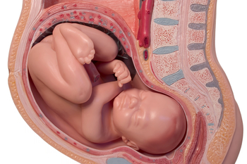
    iStock_000013877415XSmall-1-thumb-285x177-14594Brain Injury from Birth: a medical malpractice lawsuit may arise may when a child’s brain is negligently deprived of oxygen during pregnancy, labor and delivery. This may result in the child later developing cerebral palsy, mental retardation, seizures, blindness, deafness, and learning disabilities. Oxygen deprivation that injures a baby’s brain may arise from any of the following events:

    *Compression of the umbilical cord during delivery
    *Maternal Infection present during the pregnancy or delivery
    *Placental abruption or uterine rupture prior to birth
    *Maternal high blood pressure during the pregnancy (preclampsia)
    *Breeched vaginal position of the baby (feet first rather than head first)
    *Improper administration or doasage of epidural or labor inducing drugs during the delivery
    *Failure to timely perform an emergency c-section
    *Fetal macrosomia (oversized baby) unable to navigate the birth canal 

    Brain Injury in Adults and Children: a medical malpractice lawsuit may arise as a result of errors in diagnosis and treatment of a serious medical condition. A few of the causes of brain injury in children and adults that may involve medical malpractice include:

    *Medication errors
    *Anesthesia errors
    *Surgical errors
    *Radiology errors 
    *Emergency room errors
    *Delay in diagnosis/treatment of heart attack or cardiac arrest
    *Delay in diagnosis/treatment of a stroke, aneurysm, or blood clot
    *Delay in diagnosis/treatment of meningitis or encephalitis
    *Delay in diagnosis/treatment of a spreading infection or abscess
    *Delay in diagnosis/treatment of internal bleeding
    *Delay in diagnosis/treatment of hydrocephalus 
    *Delay in diagnosis/treatment of diabetes/diabetic coma/insulin shock

    In addition to the above mentioned causes of brain injury, many other errors involving patient treatment and care may give rise to a lawsuit.  One of the most common causes occurs when a hospital patient or nursing home patient falls (due to inadequate protective measures) and the patient suffers a traumatic brain injury.  In fact, falls are the leading cause of traumatic brain injury in our country surpassing even motor vehicle accidents. For a detailed guide to the incidence, prevalence, and epidemiology of brain injury, seeEssential Brain Injury Guide prepared under the auspices of the Brain Injury Association of America.     


  2. Advanced Diagnostic Imaging for Acquired Brain Injury

    Posted by Dr. Jack Sacks, Esq.on March 22, 2016

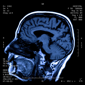
    One of the first steps in evaluating brain injury is diagnostic imaging. Imaging refers to various methods of viewing the structures and processes residing in the brain. Some of the more familiar modalities are CT (or CAT) scans, which use X-rays to evaluate intracranial structures. MRI, Magnetic Resonance Imaging, uses magnetic fields to illustrate the brain. However, in cases of traumatic brain injury (TBI), more advanced methods may be needed for proper diagnosis.

    iStock_000000113655XSmall.jpg

    An MRI machine can use special software to perform a brain scan called Diffusion Tensor Imaging (DTI). This scan detects the diffusion of water across brain cells and highlights certain areas that may be associated with injury. These injuries may not be apparent on conventional MRI’s.

    PET scan (Positron Emission Tomography) measures uptake and metabolism of glucose from a small radioactive “tagged” sample injected into the patient.  The scanner monitors this sample as the brain utilizes it. The metabolic uptake and usage may differ in patients who have suffered a brain injury. This helps define the extent and type of injury.

    In SPECT (Single Photon Emission Computed Tomography), the tagged sample is not absorbed or utilized in the brain cells. Instead, it remains in the blood stream and demonstrates the blood movement or perfusion through the brain. Areas of brain injury or damage may not have normal blood flow so the SPECT scan helps define these areas.

    Brain injuries can result from medical malpractice. A baby may suffer birth trauma during labor and delivery. A child may have a concussion or sports injury misdiagnosed or improperly evaluated by a physician. An individual injured in a car accident may not have the indicated testing done by the emergency room. Although scientific progress in imaging studies has improved the ability to diagnose and evaluate brain injuries, these tests need to be utilized in the appropriate situations so patient results and outcomes can improve.


  3. Brain Injuries and Birth

    Posted by Dr. Jack Sacks, Esq.on March 01, 2016

    The brain is responsible for higher motor and sensory functions. It requires a constant source of oxygen in order to continue its vital functions. When the flow of oxygen is interrupted, the consequences can be devastating.  Severe injury can occur when the brain lacks the oxygen needed to continue functioning. A total lack of oxygen can be referred to as “anoxic” injury whereas a partial lack of flow can be referred to as “hypoxic” injury.

    Brain injury can occur in an unborn baby if the flow of oxygen from the mother’s circulation to the baby’s is interrupted. This can happen from a number of causes. For example, the placenta can become partially detached from the wall of the uterus (placental abruption), which can interrupt the flow of oxygen to the baby. This complication can arise in pregnant patients with untreated or inadequately treated high blood pressure. Close monitoring of mother and baby, along with timely delivery and/or C-Section when needed can prevent these complications.

    iStock_000000195410Large.jpg

    Excessive contractions of the uterus (sometimes called “uterine hyperstimulation” or “tetanic contractions”) from labor, or from medicines used to accelerate labor, like pitocin, can cause problems with the baby’s oxygen supply. The rapid, powerful contractions of the uterus can prevent maternal oxygen from reaching the baby. If the flow of oxygen to the baby is interrupted, hypoxic or anoxic brain injury can occur. The consequences of this can be severe and may include seizures, brain damage, developmental delay, cerebral palsy, and other problems with motor or cognitive functions. Use of medications like pitocin must be closely monitored by dosage and effect on the mother and baby. If contractions are too strong or too frequent, the dosage may need to be decreased, the medicine may need to be stopped entirely, or special medication to reverse the effects may be given.

    Brain injuries may occur in infants, children, and adults. Stroke, cardiac arrest, or choking can all interrupt the flow of oxygen to the brain. Patients who are undergoing surgery, or are in an intensive care unit in a hospital often have their oxygen levels monitored to be sure they are getting a sufficient supply. If they are not properly monitored or complications occur, hypoxic or anoxic brain injuries may result.

    Medical malpractice that results in brain injury is devastating for the victim as well as families and loved ones. The extent of disability from hypoxic or anoxic brain injury varies greatly, but many people require extensive care and rehabilitation to regain function and improve their quality of life.


  4. Pheochromocytoma and Medical Malpractice

    Posted by Dr. Jack Sacks, Esq.on May 15, 2012

    Pheochromocytoma is an adrenal gland tumor comprised of chromaffin cells that produce and release excess epinephrine and norepinephrine, which are hormones that effect heart rate, metabolism, and blood pressure. Pheochromocytomas are generally benign and can appear at any age; however, they commonly occur during middle age.

    iStock_000017392233XSmall.jpg


    If left untreated or unrecognized, this tumor can be life threatening. Researchers have yet to discover the underlying cause of pheochromocytoma. However, certain disorders such as Multiple Endocrine Neoplasia type II (MEN II), Von Hippel Lindau disease, Neurofibromatosis 1, and Familial Paraganglioma are associated with pheochromocytoma.

    Isolated, paroxysmal episodes of hypertension occur in fewer than half of individuals with pheochromocytoma. Episodes of hypertension can occur at unpredictable intervals and usually last 15-20 minutes. During these episodes, the patient experiences hypertension, tachycardia, and fever; however, the patient’s vital signs can be normal at other times. As the tumor grows, episodes increase in frequency, length, and severity. Additional symptoms include abdominal pain, chest pain, irritability, pallor, and weight loss.

    However, two thirds of patients experience chronic sustained hypertension. Whether sustained or episodic, the patient’s hypertension is associated with an increased risk of myocardial ischemia, heart failure, renal injury, and cerebrovascular accidents. Sudden cardiac death may occur secondary to catecholamaine-induced myocardial irritability and ventricular arrhythmias.

    Diagnosis of pheochromocytoma is based on elevated levels of free catecholamines and metabolites, such as vanillymandelic acid and metanephrines, in the patient’s urine.  The following additional tests can be used to diagnose pheochromocytoma: abdominal CT scan, adrenal biopsy, catecholamines blood test, glucose blood test, metanephrine blood test, MIBG scintiscan, and MRI of the abdomen.

    The current treatment for pheochromocytoma is surgery to excise the tumor. It is essential to stabilize the patient’s blood pressure and pulse with adrenergic blocking medication prior to surgery. When the tumor cannot be removed, medication is required to manage it. The majority of patients have benign tumors that are excisable; however, 10% of tumors recur and 25% of patients still have high blood pressure after surgery.

    A medical malpractice lawsuit may arise when a patient presents with typical signs and symptoms of pheochromocytoma but the doctor fails to make the diagnosis thereby causing bodily injury, disability or death to the patient usually as a result of uncontrolled hypertension.


  5. Lumbar Puncture – Indications and Usage

    Posted by Dr. Jack Sacks, Esq.on October 07, 2011

    Lumbar puncture is a procedure performed in the lower back area, where a needle is inserted between two vertebrae to remove a sample of cerebrospinal fluid (CSF). The patient flexes his/her back to widen the spaces between the vertebrae so it is easier for the physician to access the region. The back is washed with antiseptic soap or iodine and covered with a sterile sheet. A local anesthetic is used to numb the area and then a thin hollow needle is inserted through the spinal membrane and into the spinal canal. During this portion of the procedure, the patient generally feels pressure. The CSF pressure is measured, a small amount of fluid is removed, and the pressure is measured again. The needle is removed and the site is bandaged. The total procedure takes about 45 minutes.

    Thumbnail image for Thumbnail image for iStock_000016020161XSmall.jpg

    Lumbar puncture is used to collect CSF for analysis to help in diagnosing conditions such as subarachnoid hemorrhage (typically caused by ruptured aneurysm or traumatic brain injury), meningitis (inflammation of the membranes around the brain), and cancers of the brain or spinal cord. Lumbar Puncture may also be utilized to inject anesthetic medications, chemotherapeutic drugs, contrast material, or radioactive substances into CSF.

    Lumbar Puncture may be used to identify increased or decreased CSF pressure.  Increased CSF pressure can be caused by increased intracranial pressure as is seen with traumatic brain injury, ruptured aneurysm, and sometimes hydrocephalus. On the other hand, decreased CSF pressure can be caused by spinal cord tumor, shock, fainting, or diabetic coma.  Normal CSF appears clear and colorless. When an infection is present, the CSF may look cloudy and be yellow or pink in color. Infection may be suspected if there is an increased level of white blood cells and/or protein. Increased CSF glucose indicates hyperglycemia whereas decreased CSF glucose may reflect hypoglycemia, bacterial or fungal infection, tuberculosis, or meningitis. If tumor cells are detected, the patient may have cancer in the brain, spinal cord, or CSF.  If there are increased gamma globulin levels, the patient may be suffering from multiple sclerosis, neurosyphilis, or Guillan-Barre syndrome. The analysis of the CSF by the lab and the measuring of CSF pressure recorded from the lumbar puncture help in determining a diagnosis in many serious clinical situations.