Medical Malpractice Attorneys
  • Toll Free 1-800-529-9120
  1. Brain Injury and Medical Malpractice

    Posted by Dr. Jack Sacks on May 24, 2018.

    The prevalence of brain injury in the United States is alarming as it is the second leading cause of disability in the country. Often referred to as the silent epidemic, approximately 3.17- 5.3 million Americans suffer from traumatic brain injuries, another 4.7 million have brain injuries from strokes, and another 500,000 have cerebral palsy (brain injury due to an event of oxygen deprivation). Causes of brain injury that may give rise to a medical malpractice lawsuit are further described below:

    iStock_000013877415XSmall-1-thumb-285x177-14594Brain Injury from Birth: a medical malpractice lawsuit may arise may when a child’s brain is negligently deprived of oxygen during pregnancy, labor and delivery. This may result in the child later developing cerebral palsy, mental retardation, seizures, blindness, deafness, and learning disabilities. Oxygen deprivation that injures a baby’s brain may arise from any of the following events:

    *Compression of the umbilical cord during delivery
    *Maternal Infection present during the pregnancy or delivery
    *Placental abruption or uterine rupture prior to birth
    *Maternal high blood pressure during the pregnancy (preclampsia)
    *Breeched vaginal position of the baby (feet first rather than head first)
    *Improper administration or doasage of epidural or labor inducing drugs during the delivery
    *Failure to timely perform an emergency c-section
    *Fetal macrosomia (oversized baby) unable to navigate the birth canal 

    Brain Injury in Adults and Children: a medical malpractice lawsuit may arise as a result of errors in diagnosis and treatment of a serious medical condition. A few of the causes of brain injury in children and adults that may involve medical malpractice include:

    *Medication errors
    *Anesthesia errors
    *Surgical errors
    *Radiology errors 
    *Emergency room errors
    *Delay in diagnosis/treatment of heart attack or cardiac arrest
    *Delay in diagnosis/treatment of a stroke, aneurysm, or blood clot
    *Delay in diagnosis/treatment of meningitis or encephalitis
    *Delay in diagnosis/treatment of a spreading infection or abscess
    *Delay in diagnosis/treatment of internal bleeding
    *Delay in diagnosis/treatment of hydrocephalus 
    *Delay in diagnosis/treatment of diabetes/diabetic coma/insulin shock

    In addition to the above mentioned causes of brain injury, many other errors involving patient treatment and care may give rise to a lawsuit.  One of the most common causes occurs when a hospital patient or nursing home patient falls (due to inadequate protective measures) and the patient suffers a traumatic brain injury.  In fact, falls are the leading cause of traumatic brain injury in our country surpassing even motor vehicle accidents. For a detailed guide to the incidence, prevalence, and epidemiology of brain injury, seeEssential Brain Injury Guide prepared under the auspices of the Brain Injury Association of America.     


  2. Increased Fluid Around the Heart May Cause Cardiac Tamponade

    Posted by Dr. Jack Sacks on April 30, 2018

    Pericardial effusion occurs when there is an abnormal amount of fluid around the heart.  The heart is normally surrounded by a thin membranous sac called the pericardium.  The space between the pericardium and the muscle that is the heart is referred to as the perciardial space.  Normal levels of pericardial fluid within the pericardial space are from 15 to 50 mL, or about 1-3 tablespoons.

    iStock_000003834803XSmall.jpg

    An effusion, therefore, represents  an abnormal accumulation of fluid in the pericardial space.   Because of the limited amount of space in the pericardial cavity, fluid accumulation will lead to increased intrapericardial pressure and this can negatively affect heart function.  Cardiac tamponade occurrs when there is a  large enough pericardial effusion causing enough pressure to adversely affect heart function.  This is an emergent life threatening condition.

    Pericardial effusion symptoms may include difficulty breathing (dyspnea), shortness of breath when lying down (orthopnea), chest pain, cough, dizziness, low grade fever, rapid heart rate (tachycardia), and a feeling of anxiety

    Pericardial effusion may be caused by:

    -a disturbed equilibrium between the production and re-absorption of pericardial fluid,
    -a structural abnormality that allows fluid to enter the pericardial cavity
    -inflammation of the pericardium (pericarditis)
    -bacterial or viral infections
    -injury to the heart from a medical procedure
    -cancer
    -heart attack
    -autoimmune disorders

    Unfortunately, pericardial effusion and cardiac tamponade may result from improper placement of a central venous catheter during a medical procedure involving an infant.  This may arise when there is an inadvertent perforation into the pericardial space by the CVC and fluids are artificially infused into the space thereby causing the tamponade. Upon recognition of this situation, emergent removal of this fluid via a needle inserted through the chest wall and into the pericardial space (pericardiocentesis) can improve the infant’s chance of survival. It is suggested that routine radiography be performed to readily identify the CVC tip in all cases when these lines are placed into babies.    Increased awareness of this complication may decrease the mortality associated with CVC related pericardial effusions.

    Treatment depends on the underlying cause and the severity of the heart impairment. Pericardial effusion due to a viral infection sometimes goes away within a few weeks without treatment.  Some pericardial effusions remain small and never need treatment.  If the pericardial effusion is due to an autoimmune condition treatment with anti-inflammatory medications may help. If the effusion is compromising heart function and causing cardiac tamponade, it will need to be drained, most commonlyby a pericardiocentesis.  In some cases, surgical drainage may be required by cutting through the pericardium creating what is referred to as a pericardial window.


  3. Deep Venous Thrombosis and Medical Malpractice

    Posted by Dr. Jack Sacks, Esq.on March 28, 2016

    Deep venous thrombosis is the development of a blood clot in the large, deep veins of the lower leg and thigh. Thrombi can cause tissue injury due to vascular occlusion or distal embolization. However, venous obstruction can be offset by collateral blood vessels. Thrombi can also cause local pain and edema due to the blockage of blood flow. If the clot breaks off and travels through the blood, it is referred to as an embolism. An embolism can become trapped in the brain, lungs, or heart leading to major injury. Pulmonary embolus (PE) is a common complication and life threatening if not treated quickly with anticoagulants. PE presents with shortness of breath, chest pain, and cough with blood in sputum.

    iStock_000012053156XSmall.jpgDVT can occur with stasis or in hypercoaguable states. It is commonly seen following trauma, surgery, or burns, which contribute to decreased physical activity, damage to vessels, and release of procoagulant substances from tissues. Reduced physical activity causes a decline in the milking action of lower leg muscles and slows venous return. Risk factors for DVT include advanced age, bed rest, immobilization, smoking, birth control pills, family history of blood clots, fractures in the pelvis or legs, giving birth within the last 6 months, heart failure, and obesity.  To prevent DVT, patients should move their legs during long flights or when they are immobile for long periods of time.

    Although many DVTs are asymptomatic, they can recur. Some individuals suffer from post-phlebitic syndrome, which involves chronic pain and swelling in the leg. The major symptoms of DVT include changes in a patient’s leg such as redness, increased temperature, pain, and tenderness. Diagnosis is based on the physical exam, which should demonstrate a red, swollen leg. Diagnostic tests include a D-dimer blood test along with other blood tests to check for hypercoagulability such as activated protein C resistance, anti-thrombin III levels, antiphospholipid antibodies, and genetic testing for mutations with a predisposition towards blood clots. Imaging studies of the legs include Doppler ultrasound, plethysmography, and radiography.

    The primary treatment for DVT is anti-coagulants, also known as blood thinners. They prevent the formation of new clots and the growth of old clots. However, they cannot dissolve existing clots. Patients are more likely to bleed on these medications. Heparin is an IV administered anticoagulant given in a hospital setting.  Warfarin (Coumadin) is an oral anticoagulant that takes several days to work; thus, Heparin cannot be stopped until Warfarin is functioning at an effective dose for a minimum of two days. Many patients wear pressure stockings on their legs to improve blood flow and decrease their risk of DVT.  When medications are ineffective, patients may need to undergo surgery. A filter can be placed in the body’s largest vein to prevent thrombi from migrating to the lungs. Also, surgery may be necessary to remove large thrombi.


  4. Meningitis and Medical Malpractice

    Posted by Dr. Jack Sacks, Esq.on March 28, 2016

    Meningitis is inflammation of the meninges, the membranes that cover the brain and spinal cord. It is commonly caused by infection, but other causes include chemical irritants, drug allergies, fungi, and tumors. Based on the clinical evolution of the illness and the type of inflammatory exudate present in the cerebral spinal fluid (CSF), infectious meningitis is categorized into acute pyogenic (usually bacterial), aseptic (usually viral), and chronic (usually tuberculosis, spirochetal, cryptococcal).

    iStock_000003651637XSmall.jpg

    Early diagnosis is essential for bacterial meningitis because it can result in death or brain damage if left untreated. In bacterial meningitis, a correlation exists between bacterial organism and age. The most likely organism in neonates may be Escherichia Coli or group B Streptococci. In the elderly, it may be Streptococcus Pneumonia or Listeria Monocytogenes. In young adults, it may be Neisseria Meningitides. In contrast, most viral infections are due to enteroviruses but only a small number of people who develop enteroviral infections present with meningitis. Other viral infections that can cause meningitis include mumps, herpes virus, measles, and influenza. Chronic meningitis can be caused by pathogens such as mycobacteria and spirochetes. Thus, medical attention is necessary to differentiate between bacterial, viral, and chronic meningitis.

    Risk factors include individuals over the age of 60 or below the age of 5, diabetes mellitus, renal or adrenal insufficiency, hypoparathyroidism, cystic fibrosis, immunosuppression, HIV, crowding (military recruits and college residents), recent exposure to those with meningitis, etc. The symptoms have a rapid onset and include fever, chills, mental status changes, nausea, vomiting, photophobia, severe headache, and meningismus (stiff neck). Additional symptoms include agitation, bulging fontanelles, decreased consciousness, tachypnea, poor feeding or irritability in children, and opisthotonos (unusual posture, with head and neck arched backwards).

    To confirm a diagnosis, a lumbar puncture (spinal tap) should generally be performed on anyone suspected of meningitis to sample and culture the CSF for abnormal cell counts, glucose, and protein. Other diagnostic tests include blood culture, chest x-ray, and MRI or CT scan of the head. The underlying cause of the meningitis needs to be determined to administer proper treatment and define the severity of each case. Unlike bacterial meningitis, viral meningitis usually does not involve treatment and patients generally recover within two weeks; however, in certain instances (such as with the herpes simplex virus) antiviral medications may be indicated.

    Antibiotic treatment for bacterial meningitis is dependent on the underlying bacterium. By treating the most common types, the risk of dying is reduced to below 15%. Symptoms such as brain swelling, shock, and seizures are treated with other medications and intravenous fluids. Possible complications of meningitis include brain damage, subdural effusion, hearing loss, hydrocephalus, and seizures. To prevent contraction of meningitis, the meningococcal vaccination is recommended for populations at risk.


  5. Advanced Diagnostic Imaging for Acquired Brain Injury

    Posted by Dr. Jack Sacks, Esq.on March 22, 2016

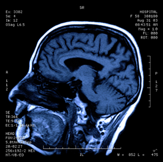
    One of the first steps in evaluating brain injury is diagnostic imaging. Imaging refers to various methods of viewing the structures and processes residing in the brain. Some of the more familiar modalities are CT (or CAT) scans, which use X-rays to evaluate intracranial structures. MRI, Magnetic Resonance Imaging, uses magnetic fields to illustrate the brain. However, in cases of traumatic brain injury (TBI), more advanced methods may be needed for proper diagnosis.

    iStock_000000113655XSmall.jpg

    An MRI machine can use special software to perform a brain scan called Diffusion Tensor Imaging (DTI). This scan detects the diffusion of water across brain cells and highlights certain areas that may be associated with injury. These injuries may not be apparent on conventional MRI’s.

    PET scan (Positron Emission Tomography) measures uptake and metabolism of glucose from a small radioactive “tagged” sample injected into the patient.  The scanner monitors this sample as the brain utilizes it. The metabolic uptake and usage may differ in patients who have suffered a brain injury. This helps define the extent and type of injury.

    In SPECT (Single Photon Emission Computed Tomography), the tagged sample is not absorbed or utilized in the brain cells. Instead, it remains in the blood stream and demonstrates the blood movement or perfusion through the brain. Areas of brain injury or damage may not have normal blood flow so the SPECT scan helps define these areas.

    Brain injuries can result from medical malpractice. A baby may suffer birth trauma during labor and delivery. A child may have a concussion or sports injury misdiagnosed or improperly evaluated by a physician. An individual injured in a car accident may not have the indicated testing done by the emergency room. Although scientific progress in imaging studies has improved the ability to diagnose and evaluate brain injuries, these tests need to be utilized in the appropriate situations so patient results and outcomes can improve.


  6. Spinal Cord Compression and Cauda Equina Syndrome

    Posted by Dr. Jack Sacks, Esq.on January 03, 2016

    There are approximately 12,000 new cases of spinal cord compression annually in the United States with the average age of victims being 39.5 years old. Causes of spinal cord compression include trauma (such as auto accidents, falls, sports injury, epidural injection), spinal abscess, tumor, hematoma or blood clot, ruptured or herniated disk, and spinal stenosis. . Early symptoms may include the start of loss of movement or feeling in the arms or legs, back pain, and the loss of bowel or bladder function or control. Spinal cord compression of sudden onset constitutes a medical emergency, as the longer the duration of symptoms before cord pressure relief is obtained by surgical intervention (laminectomy), the greater the chance of permanent injury. Diagnosis is by clinical exam and other testing such as X-ray, CT, and MRI. MRI is usually the most accurate study to detect spinal cord compression as details of both bony and soft tissue abnormalities in the spinal column may be visualized. MRI may also be the preferred study if spinal cord injury occurs during pregnancy as MRI offers reduced radiation exposure to the fetus. However, after an accident, traction devices to immobilize the spine and life support equipment may preclude the use of MRI.

    The spinal column is comprised in descending order of the cervical, thoracic, lumbar, and sacral areas. The actual spinal cord ends at the level of L-1. Below the level of L-1, the cord branches into a bundle of spinal nerve roots from L-1 through L-5 and S-1 through S-5 that resembles a horses tail referred to as the cauda equina. When the nerve roots of the cauda equina are compressed permanent neurogenic injury may occur. This may be referred to as cauda equina syndrome (CES). Causes of compression of these nerve roots include the same types of causes as listed above for spinal cord compression. Symptoms of early onset of cauda equina syndrome include numbness in the groin (saddle anesthesia), loss of bowel or bladder function or control, weakness in the legs, and absence of ankle reflexes. Early diagnosis and treatment of CES is important as the longer the duration of symptoms, the more likely permanent neurological injury will occur such as paralysis and incontinence. Cauda equina syndrome of sudden onset is a medical emergency and treatment generally involves surgical decompression of the affected nerve roots. A laminectomy may be performed to relieve pressure on the nerve root in cases where there is a herniated or ruptured disk, hematoma, abscess, or tumor.

    In spinal cord and nerve root compression cases involving sudden onset of neurological symptoms, medical malpractice may arise when there has been an unreasonable delay in diagnosis and prompt surgical intervention by the doctors or hospital. Allegations of medical malpractice may also include a failure to diagnose and treat a vertebral fracture of the neck or back that required traction or immobilization which was not performed, thereby allowing pathological movement that resulted in spinal cord compression.


  7. Septic Arthritis and Medical Malpractice

    Posted by Dr. Jack Sacks, Esq.on December 31, 2011

    Septic arthritis, also known as reactive arthritis and bacterial arthritis, is inflammation of a joint caused by infection. When it develops, it represents a medical emergency. It develops when bacteria or fungus is transmitted through an individual’s bloodstream to a joint. Individuals can develop septic arthritis at any age but it is less common between the ages of three to adolescence.

    The majority of acute cases involve bacteria such as staphylococcus or streptococcus. However, children who develop septic arthritis tend to be infected with Group B streptococcus or Haemophilus influenza, if they have not been vaccinated. The number of cases that progress to a chronic infection are less common. Chronic infections are generally caused by organisms such as Mycobacterium tuberculosis and Candida albicans.

    Overall, the most common sites of infection are the hip and knee joints. The following are considered risk factors for septic arthritis: artificial joint implants, bacterial infection, chronic disease (diabetes, rheumatoid arthritis, and sickle cell disease), intravenous drug use, immunosuppressive medications, and recent joint surgery or arthroscopy.

    Septic arthritis generally presents with a rapid onset of symptoms. Patients develop a fever in addition to joint swelling, redness, and pain in the infected joint. Patients may also experience pseudoparalysis, which is an inability to move the extremity possessing the infected joint. To diagnose septic arthritis, physicians will aspirate the joint fluid to check the cell count, look for crystals under the microscope, and perform a gram stain and culture. In addition, other tests include a blood culture and an X-ray of the affected joint.

    Possible complications include joint degeneration and permanent joint damage, which can occur if appropriate treatment is delayed. If antibiotics are promptly given to treat the infection the patient has a good prognosis. If fluid accumulates in the joint, aspiration may be necessary. Aspiration involves a needle being inserted into the joint to drain off the excess fluid. Only severe cases require surgery to remedy the infected joint fluid; however, if a prosthetic joint is affected, it may need to be replaced.


  8. Pituitary Adenomas and Medical Malpractice

    Posted by Dr. Jack Sacks, Esq.on October 25, 2011

    A pituitary adenoma is generally a benign, slow growing tumor that occurs in the pituitary gland. The pituitary gland is a small, bean shaped structure that lies at the base of the brain. It has a central role in the regulation of hormones that affect the body such as Adrenocorticotropic hormone (ACTH), Growth hormone (GH), Prolactin, and Thyroid-stimulating hormone (TSH).

    Approximately 1 in 1,000 individuals have pituitary adenomas. They are generally not cancerous but may invade nearby structures.  They are classified based on size. A microadenoma is less than 1 cm in diameter whereas a macroadenoma is larger than 1 cm in size.

    iStock_000017548218XSmall.jpg

    Based on whether the pituitary adenoma is a hormone-producing or hormone-inactive tumor, the patient will present with different symptoms. Hormone-producing tumors will make excessive amounts of an active hormone so symptoms present as a hormonal imbalance. The three most common hormone-producing adenomas are Prolactinomas, Growth hormone-secreting pituitary adenoma, and ACTH-secreting pituitary adenoma.

    If the patient has a large hormone-inactive or hormone-producing tumor, it may compress surrounding brain structures due to its size.  Large pituitary tumors may compress the pituitary gland contributing to pituitary failure, which can lead to sexual dysfunction, inadequate body cortisol levels, and hypothyroidism. Other possible presentations relating to compression of brain structures include visual loss, headache, the “stalk effect”, and pituitary apoplexy.  The “stalk effect” is due to the compression of the pituitary stalk, the structure connecting the brain to the pituitary gland, which leads to a mild elevation in the hormone prolactin. Higher levels of prolactin in females can contribute to irregular menstrual cycles. Pituitary apoplexy presents with an abrupt headache and visual loss. It can occur under two different situations. The pituitary adenoma can bleed internally causing a sudden increase in size or the tumor can outgrow its blood supply and the dead tissue will swell.

    Pituitary adenomas can be diagnosed based on endocrine function testing, imaging, and visual field testing. Often times, visual problems may be the only symptom present.  Optometrists and ophthalmologists should seriously consider the possibility of a pituitary tumor when the patient presents with an unexplained loss of visual field (especially peripheral vision), double vision, or blurred vision as the growth of a pituitary tumor can cause compression on the visual pathway (retina, optic nerve, etc). A delay in diagnosis of a pituitary adenoma may result in permanent loss of vision due to the damage caused by this compression. Endocrine function testing evaluates cortisol, follicle-stimulating hormone, lutenizing hormone, insulin growth factor-1, prolactin, testosterone/estradiol, and thyroid hormone levels. The preferred method of imaging is MRI, which screens for adenomas larger than 4 mm.

    Treatment for pituitary adenomas depends on the presence of hormone production, size of the tumor, invasion of the tumor into surrounding structures, and the age and health of the patient. Drug therapy is used to treat hormone-producing tumors. For example, bromocriptine and cabergoline are used to treat tumors secreting prolactin because these medications decrease prolactin levels and tumor size. Pituitary adenomas that require surgery are usually minimally invasive techniques, where the tumor is removed through the nose.  On the other hand, radiation therapy involves high doses of radiation being delivered to the tumor. It is a treatment utilized for pituitary adenomas that cannot be controlled by drug therapy or surgical intervention. Patients have the best outlook when the entire tumor can be removed.


  9. Lumbar Puncture – Indications and Usage

    Posted by Dr. Jack Sacks, Esq.on October 07, 2011

    Lumbar puncture is a procedure performed in the lower back area, where a needle is inserted between two vertebrae to remove a sample of cerebrospinal fluid (CSF). The patient flexes his/her back to widen the spaces between the vertebrae so it is easier for the physician to access the region. The back is washed with antiseptic soap or iodine and covered with a sterile sheet. A local anesthetic is used to numb the area and then a thin hollow needle is inserted through the spinal membrane and into the spinal canal. During this portion of the procedure, the patient generally feels pressure. The CSF pressure is measured, a small amount of fluid is removed, and the pressure is measured again. The needle is removed and the site is bandaged. The total procedure takes about 45 minutes.

    Thumbnail image for Thumbnail image for iStock_000016020161XSmall.jpg

    Lumbar puncture is used to collect CSF for analysis to help in diagnosing conditions such as subarachnoid hemorrhage (typically caused by ruptured aneurysm or traumatic brain injury), meningitis (inflammation of the membranes around the brain), and cancers of the brain or spinal cord. Lumbar Puncture may also be utilized to inject anesthetic medications, chemotherapeutic drugs, contrast material, or radioactive substances into CSF.

    Lumbar Puncture may be used to identify increased or decreased CSF pressure.  Increased CSF pressure can be caused by increased intracranial pressure as is seen with traumatic brain injury, ruptured aneurysm, and sometimes hydrocephalus. On the other hand, decreased CSF pressure can be caused by spinal cord tumor, shock, fainting, or diabetic coma.  Normal CSF appears clear and colorless. When an infection is present, the CSF may look cloudy and be yellow or pink in color. Infection may be suspected if there is an increased level of white blood cells and/or protein. Increased CSF glucose indicates hyperglycemia whereas decreased CSF glucose may reflect hypoglycemia, bacterial or fungal infection, tuberculosis, or meningitis. If tumor cells are detected, the patient may have cancer in the brain, spinal cord, or CSF.  If there are increased gamma globulin levels, the patient may be suffering from multiple sclerosis, neurosyphilis, or Guillan-Barre syndrome. The analysis of the CSF by the lab and the measuring of CSF pressure recorded from the lumbar puncture help in determining a diagnosis in many serious clinical situations.