Medical Malpractice Attorneys
  • Toll Free 1-800-529-9120
  1. Oral Cancer and Medical Malpractice

    Posted by Dr. Jack Sacks, Esq.on March 26, 2016

    Oral cancer is the uncontrollable growth of cells that invade and inflict damage in areas involving the lips, tongue, cheek lining, floor of the mouth, gingiva, and palate. Men are twice as likely to have oral cancer, specifically men over the age of 40. Risk factors include smoking/tobacco use; heavy alcohol use; chronic irritation from rough teeth, dentures, or fillings; human papilloma virus infection; family history; excessive sun exposure; taking immunosuppressants that weaken the immune system; and poor oral hygiene. Oral cancer can present with a sore, lump, or ulcer in the mouth that initially is painless; however, as the cancer progresses it may evolve into a burning sensation or pain. The area may appear pale colored or it can be dark and discolored. Other symptoms include dysphagia (difficulty swallowing), odynophagia (painful swallowing), chewing problems, speech difficulties, lymphadenopathy (swollen lymph nodes), and weight loss.

    iStock_000018882292XSmall.jpg

    To diagnose oral cancer, your physician or dentist will examine your oral cavity and if a suspicious lesion is identified then an oral brush biopsy may be performed. The test is painless and involves isolating and analyzing a small sample of tissue for abnormal cells. However, if the lesion is more concerning then a scapel biopsy is recommended to determine whether the area is malignant or benign. X-rays and CT scans may be utilized to determine if the cancer has metastasized. Other tests that may be conducted include endoscopy, barium swallow, or PET scan. 90% of oral cancers are squamous cell carcinomas. Squamous cells are thin, flat cells that line the lips and oral cavity. Squamous cell carcinoma often develops in areas of leukoplakia, white patches of cells that do not rub off. Other types of oral cancer include adenocarcinoma, lymphoma, melanoma, or teratoma.

    If oral cancer is not diagnosed early, it can be life threatening. Treatment is dependent on the stage of the cancer which is determines by tumor size, lymph node involvement, and metastatsis. Surgery is recommended if the tumor is small enough and has not spread to the lymph nodes. Complications of surgery include disfigurement of the face, head, and neck. Often surgery is combined with radiation. Complications of radiation include dry mouth and dysphagia. When dealing with larger tumors, chemotherapy is recommended. Speech therapy is also essential to improve and retain movement, chewing, swallowing, and speech.

    Depending on the presentation of the oral cancer, approximately 50% of individuals with oral cancer may survive greater than 5 years following diagnosis and treatment. If the cancer is identified early before significant metastasis, then the cure rate may be almost 90%; nevertheless, the majority of oral cancers have metastasized prior to diagnosis. One in four individuals with oral cancer will die due to delayed diagnosis and treatment. To prevent oral cancer, individuals should avoid smoking/tobacco, moderate or avoid alcohol use, and practice good oral hygiene.


  2. Stomach Cancer and Medical Malpractice

    Posted by Dr. Jack Sacks, Esq.on February 28, 2016

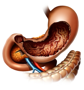
    Stomach cancer is referred to as gastric cancer. The most common type of gastric cancer is called adenocarcinoma. This starts in the glandular tissue that composes the lining of the stomach and accounts for 90% to 95% of all gastric cancers. Other forms of gastric cancer include lymphomas, which involve the lymphatic system and sarcomas, which involve the connective tissue (such as muscle, fat, or blood vessels).

    iStock_000002366269XSmall.jpg

    Generally, the best chance for cure of gastric cancer is when it is found and treated at a very early stage. Unfortunately, the outlook is typically poor if the cancer is already at an advanced stage when discovered.

    Gastric cancer can develop in any part of the stomach and may spread throughout the stomach and to other organs, such as the esophagus, liver and lungs.  Gastric cancer is responsible for about 800,000 deaths worldwide per year.

    The signs/symptoms include indigestion and/or heartburn, loss of appetite, especially for meat, abdominal discomfort or irritation, weakness and fatigue, bloating of the stomach, usually after meals, abdominal pain in the upper abdomen, nausea and occasional vomiting, diarrhra or constipation, weight loss, vomiting, blood in the stool, which will appear as black. This can lead to anemia, and dyspagia (trouble swallowing), which suggests a tumor in the upper protion of the stomach or extension of the gastric tumor into the esophagus.

    The diagnosis is typically made when abnormal tissue seen in a direct visual (gastroscopic) examination is biopsied. This tissue is then examined under a microscope to check for the presence of cancerous cells. A biopsy, with subsequent histological analysis, is the only sure way to confirm the presence of cancer cells.

    The clinical stages of stomach cancer are:

    Stage 0. Limited to the inner lining of the stomach.

    Stage I. Penetration to the second or third layers of the stomach (Stage 1A) or to the second layer and  nearby lymph nodes (Stage 1B).

    Stage II. Penetration to the second layer and more distant lymph nodes, or the third layer and only nearby lymph nodes, or all four layers but not the lymph nodes.

    Stage III. Penetration to the third layer and more distant lymph nodes, or penetration to the fourth layer and either nearby tissues or nearby or more distant lymph nodes.

    Stage IV. Cancer has spread to nearby tissues and more distant lymph nodes, or has metastatized to other organs.

    Treatment for gastric cancer depends on both the tissue type and the stage of the cancer. Treatment for adenocarcinoma may include:

    1. Surgery– the goal of surgery is to remove all of the cancer and allow for a margin of healthy tissue,  when possible. The surgeon also removes lymph nodes surrounding the stomach to determine if they  have been invaded by cancer cells. Removing part of the stomach may relieve signs and symptoms of a  growing tumor in people with advanced stomach cancer. In this case, surgery generally does not cure the  cancer, but it can make the patient more comfortable. This is known as palliative therapy.

    2. Radiation Therapy-uses high-powered beams of energy to kill cancer cells. It can be used before surgery (neoadjuvant radiation) to shrink a stomach tumor so it’s more easily removed. Radiation therapy can also be used after surgery (adjuvant radiation) to kill any cancer cells that might remain.  Radiation is often combined with chemotherapy. In cases of advanced cancer, radiation therapy may be  used to relieve side effects caused by a large tumor. Radiation therapy can cause diarrhea, indigestion,  nausea and vomiting.

    3. Chemotherapy-is a drug treatment that uses chemicals to kill cancer cells. Chemotherapy drugs can  kill cancer cells that may have spread beyond the stomach. Chemotherapy can be given before surgery  (neoadjuvant chemotherapy) to help shrink a tumor so it can be more easily removed. And it can be  given after surgery (adjuvant chemotherapy) to kill any cancer cells that might remain in the body.    Chemotherapy is often combined with radiation therapy. Chemotherapy may be used alone in patients  with advanced stomach cancer to help relieve signs and symptoms. Side effects of chemotherapy depend  on which drugs are used, and which drugs are used depends upon the type of cancer being treated.

    4. Clinical Trials-are always being done at research cancer centers to study new treatments and new ways of using existing treatments. Participating in a clinical trial may give a patient a chance to try the  latest treatments. In some cases, researchers might not be certain of a new treatment’s side effects.

    Depending on the stage at diagnosis, the average 5 year survival rates for adenocarcinoma of the stomach are as follows:

    Stage Ia – 71%

    Stage Ib – 57%

    Stage IIa – 45%

    Stage IIb – 33%

    Stage IIIa – 20%

    Stage IIIb – 14%

    Stage IV – Less than 4%.

    Medical malpractice may be seen when there is a significant and negligent delay in the diagnosis of gastric cancer. As the above shows, if the diagnosis is delayed, and the stage advances, the prognosis for 5 year survival rates becomes poor.  During patient screening, a doctor should be aware of the risk factors for gastric cancer that include smoking cigarettes, a diet high in salted, smoked, pickled meats or foods, stomach inflammation, a history of Helicobacter pylori infection, gastric polyps, pernicious anemia, and a family history of one or more relatives diagnosed with gastric cancer.  Screening for one type of adenocarcinoma called HDGC or Hereditary Diffuse Gastric Cancer includes genetic testing for the CDH1 gene mutation.  In families with the CDH1 gene mutation, intense surveillance may be appropriate and in some cases prophylactic gastrectomy.

    Aside from upper endoscopy, other helpful diagnostic tests for gastric cancers include a double contrast barium x-ray, ultrasound, or a CT scan; however, a negative barium x-ray study alone may give false assurance and is sometimes a reason for a delay in diagnosis. Research suggests that improvement in five-year survival requires not only improved awareness by patients, but especially improved diagnostic methods and screening programs by their doctors.


  3. BREAST CANCER AND MEDICAL MALPRACTICE

    Posted by Dr. Jack Sacks, Esq.on October 18, 2015

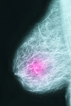
    Breast cancer affects millions of women in America and some studies indicate up to one out of eight women will be diagnosed with breast cancer over the course of her lifetime. The survival rate of breast cancer that is detected and treated early is much better than when a malignant tumor is found after it hasalready spread to lymph nodes or surrounding tissue.  Therefore, it is important that every effort be made to diagnose and treat breast lesions as quickly as possible.iStock_000013080813XSmall-thumb-300x400-8870-thumb-300x400-8871-thumb-300x400-8872.jpg

    One of the mainstays in breast cancer detection is mammography. A mammogram involves an X-ray taken of the breasts, usually taken from several angles. Some abnormalities on mammograms such as masses or calcifications can alert the healthcare provider and patient that additional diagnostic testing needs to be done. The mammogram is one of the main methods of screening for breast cancer. Other imaging methods are less commonly used for screening, such as sonography (ultrasound), and MRI. However, these diagnostic methods can be used to add to information already obtained from a mammogram study when a patient is evaluated and uncertainty or an abnormality exists.

    Patients need to be informed about the need for breast cancer screening. The mammogram, ultrasound, or MRI tests need to be read and interpreted correctly in order to diagnose problems and treat them early. In the case of breast cancer, early detection and diagnosis is key to improved cure rates.  Special circumstances and risk factors (such as family history) may point to additional diagnostic testing to help identify breast cancer in those women at high risk. If tests are not interpreted correctly, or the results are not acted on in a timely or proper fashion by the healthcare provider, medical malpractice may be the result.

    Sometimes patients or healthcare providers feel a breast mass or lump on examination. This often requires further evaluation usually beyond just a “regular” screening mammogram. As noted above, additional mammogram views, ultrasound, and/or MRI can help characterize the type of mass. However, breast masses usually warrant a visit to a surgeon or breast specialist to evaluate the patient for biopsy. Breast biopsies can be much less invasive to patients than in years past. During a “stereotactic” breast biopsy, the doctor employs mammography to locate the specific abnormal area and then a special instrument is used to remove cells from that precise area. Ultrasound can also be used to identify an abnormal area that is then biopsied. These methods are particularly useful when a breast mass cannot be felt. Sometimes when a mass can be felt, or palpated, the biopsy can be done without using X-ray or ultrasound localization.

    Breast cancer screening can help identify women with breast cancer, as well as pre-cancerous lesions and benign breast conditions. Medical malpractice may occur as a consequence of failure to adequately evaluate, identify, screen, or diagnose breast cancer. Allegations of medical malpractice may include a failure by the internist to refer the patient to a breast specialist or surgeon for evaluation, failure by the radiologist to properly read a mammogram, ultrasound, or MRI, and/or failure by the pathologist to detect cancer in a breast biopsy that is indeed malignant. With improved methods of screening, diagnosis, and treatment of breast cancer, survival rates should continue to improve.


  4. Triple Negative Breast Cancer and Medical Malpractice

    Posted by Dr. Jack Sacks, Esq.on January 31, 2012

    By dividing breast cancer into molecular breast cancer subtypes, physicians and researchers can devise better approaches and treatments for dealing with the disease. Most studies divide breast cancer into four major molecular subtypes: Luminal A, Luminal B, Triple-negative, and HER2/neu. Triple-negative breast cancer is characterized by tumors that lack estrogen, progesterone, and human epidermal growth factor receptors.

    iStock_000018505784XSmall.jpg

    Higher rates of triple negative breast cancer have been recorded in younger women, Hispanic and African American women, and women of lower socioeconomic class. Triple negative cancer tends to have a poorer prognosis than other types of breast cancer that have more available and effective treatments.

    Studies have demonstrated that triple-negative breast cancer possesses a higher probability for recurrence and spreading beyond the breast. This risk is greater within the first few years post-treatment but over time the risk becomes similar to other types of breast cancer. In addition, triple-negative tends to present as a higher grade cancer, meaning that a greater number of cancer cells are abnormal in appearance.

    Although surgical and radiation treatments are similar for different types of breast cancer, drug treatments usually vary. Many therapies for breast cancer target cellular receptors. A receptor is a molecule generally located on the surface of cells that is involved with chemical signaling. When a molecule binds to a receptor, it causes the cell to perform certain actions such as division or multiplication. Targeted drug treatments like tamoxifen or Herceptin, designed to treat HER2 positive breast cancer, have not been developed for triple negative. A patient with triple-negative does not express estrogen, progesterone, and HER2/neu receptors so therapy against these receptors is ineffective.

    Standard treatment for triple-negative breast cancer is chemotherapy. Current treatments include anthracylines, taxanes, ixabepiplone, platinum agents, and biologic agents. Patients undergoing chemotherapy can experience side effects such as vomiting, nausea, alopecia, mucositis, myelosuppression, etc. Surgery and radiation are additional avenues of treatment. Currently, cancer experts are studying several promising drug strategies targeted for triple negative breast cancer such as PARP inhibitors.