Medical Malpractice Attorneys
  • Toll Free 1-800-529-9120
  1. Small Bowel Obstruction and Medical Malpractice

    Posted by Dr. Larry Leichter on April 15, 2018

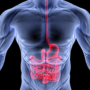
    The small bowel is a long coiled hollow tube, called a tract, that is approximately twenty-five feet long. It includes the duodenum, jejunum and ileum.  A small bowel obstruction, also known as a small intestinal obstruction, is a mechanical or functional (paralytic) blockage of the intestinal tract, which prevents the normal transit of digestive products. It can occur at any level throughout the jejunum and ileum, and is considered a medical emergency when it occurs. The condition is often treated conservatively for the first several days; however, the patient must be monitored very closely for signs of clinical deterioration that can become life threatening.
    iStock_000017493981XSmall.jpg


    Mechanical obstruction is due to a mechanical barrier, such as an adhesive band from prior surgery, which creates a road block to the bowel.  On the other hand, functional obstruction is caused by an event that interferes with the nervous innervation of the bowel, such as electrolyte imbalances and metabolic disturbances. Functional bowel obstruction can be caused by a multitude of conditions whereas mechanical SBO is generally credited to a luminal, mural, or extra-mural mechanical barrier. A clinical syndrome exists called small intestinal pseudo-obstruction, which is characterized by manifestations of mechanical bowel obstruction in the absence of an obstructive lesion.

    The symptoms of a mechanical small bowel obstruction include abdominal fullness and/or excessive gas, abdominal distention, pains and cramps in the stomach area (specifically the mid abdomen), vomiting, constipation (inability to pass gas or stool), diarrhea, and bad breath. Acute functional small bowel dilatation is referred to as adynamic or paralytic ileus. The symptoms of paralytic obstruction, in reference to the ileus, are abdominal fullness and/or excessive gas, abdominal distention, and vomiting after eating.  The pain less closely resembles the colicky type seen in mechanical obstruction, but may be just as severe.

    The diagnosis is determined by listening to the abdomen with a stethoscope. High-pitched, tinny and clanking sounds can be heard at the onset of mechanical obstruction.  If the blockage persists for too long or the bowel is significantly damaged, due to the stretching of the blood vessels supplying it thereby decreasing blood flow, bowel sounds will decrease and eventually become silent.  The hallmark of paralytic ileus is decreased or absent bowel sounds, which can create confusion in relation to the issue of etiology if this occurs.Diagnostic tests that demonstrate obstruction include plain radiographic film of the abdomen (usually in the flat and upright position), CT scan, barium enema and upper GI series with small bowel follow through.

    Treatment depends on the cause of the obstruction. In some cases, drastic measures are necessary to save a person’s life, while in others a strategy of watchful waiting is more appropriate. In general, more serious cases that require immediate treatment can be identified based on a patient’s vital signs and physical exam. If the person is very sick and appears to be on the brink of a serious event, surgery may be required to ensure the patient’s life.

    To determine if there is any deterioration consistent with lack of blood flow, which leads to bowel ischemia, gangrene, perforation, septic shock, and death, it is imperative that the following steps be taken. The bowel must be decompressed with a long indwelling tube, all oral feeding must be stopped and IV therapy must be initiated with continuous monitoring and observation. Generally speaking, there is no reason anyone presenting to the emergency room with a small bowel obstruction should die in the hospital unless there are extenuating circumstances.

  2. Appendicitis: Early Diagnosis and Treatment are Essential

    Posted by Dr. Jack Sacks, Esq.on December 01, 2015

    Appendicitis is a medical emergency that requires immediate surgery to remove the appendix.  If left untreated, an inflamed appendix will eventually rupture.  When this happens intestinal contents (stool and bacteria) spill into the abdominal cavity causing infectious peritonitis, a serious and toxic inflammation of the abdominal cavity’s lining (the peritoneum).  This condition can be fatal unless it is treated quickly with surgery and strong antibiotics.

    SYMPTOMS: Usually pain is the first symptom, starting in the mid abdomen around the navel, and except in children below 3 years old, the pain tends to localize in the right lower quadrant within a few hours.  The abdominal wall becomes sensitive to gentle pressure, and the pain can be elicited through various tests the physician will use to bring it out.  One such sign is when the abdomen is gently pushed down and quickly released.  This is known as rebound tenderness and is a clinical sign the peritoneum is inflamed.  If the appendix is located beneath the cecum (first part of the colon), it may fail to elicit tenderness (silent appendix).  And if the appendix lies entirely within the pelvis, the region below the addomen, there could be a complete absence of the abdominal signs and symptoms. In such cases, a digital rectal exam will cause discomfort localized to the region of the appendix in the right lower quadrant.  Also, if the abdomen on palpation is rigid, which is known as involuntary guarding, there should be a strong suspicion of peritonitis requiring urgent surgical intervention.  The physician can perform certain other maneuvers, such as bending and rotating the right hip, and extending the hip in the prone position, which will bring about pain consistent with inflammation caused by appendicitis.  The next symptoms usually experienced are naussea and vomiting, as well as constipation.  Eventually as the inflammation progresses, fever will occur.

    BLOOD TESTS: When appendicitis is suspected, blood tests such as a CBC need to be done to try to confirm the diagnosis.  More than 80% of adults with appendicitis have a white blood cell count greater than 10,500 cells/mm3.  Another blood test commonly used is the determination of C-reactive protein (CRP),an acute-phase reactant synthesized by the liver in response to infection or inflammation.

    DIAGNOSTIC IMAGING: The plain film of the abdomen, known as a KUB is typically taken. Visualization of an appendicolith (a white colored defect in the right lower quadrant) in a patient with symptoms consistent with appendicitis is highly suggestive of appendicitis, but this occurs in fewer than 10% of cases. Another X-ray that may be utilized is the single-contrast Barium enema, which can be performed on an unprepared bowel. Absent or incomplete filling of the appendix with contrast barium coupled with pressure effect or spasm in the cecum suggests appendicitis. Though cheap in cost, once thick barium contrast is instilled, other more definitive tests will not be able to be performed.

    The appendix may be evaluated via transabdominal sonography. Many physicians believe that ultrasonography should be the initial imaging test in pregnant women and in children due to its safety. Abdominal CT scanning has become the most important imaging study in the evaluation of patients with appendicitis.  Advantages of CT scanning include its superior accuracy when compared with other imaging techniques, as well as its ready availability, noninvasiveness, and potential to reveal alternative diagnoses. MRI plays a relatively limited role in the evaluation of appendicitis due to high cost, long scan times, and limited availability.  The lack of dangerous radiation exposure makes it an attractive modality in pregnant patients where ultrasound is not helpful in making a diagnosis.

    In today’s day and age, the diagnosis of appendicitis should generally not be missed by an emergency room physician.  If in doubt a surgeon should be called to evaluate and remove the appendix, which can be done laparoscopically, so long as it remains locally inflamed or walled off, and before there is free perforation into the abdominal cavity.  A delay in diagnosis can result in lifelong complications affecting bowel function due to the scarring of the peritoneal lining.


  3. Sigmoid Colon Volvulus and Medical Malpractice

    Posted by Dr. Jack Sacks, Esq.on February 03, 2012

    The term volvulus is derived from the Latin word volve, which means to twist. A colonic volvulus occurs when a part of the colon twists on its mesentery, resulting in acute, subacute, or chronic colonic obstruction.  Volvulus involving the sigmoid colon is the most common, occuring in 75% of cases.  Sigmoid volvulus occurs when the last part of the large bowel just before the rectum (named for its “S” shape) twists on its self.  It is common in elderly men, but it is likely to occur in anyone with a redundant sigmoid colon.

    More than 60-70% of patients present with acute symptoms; the remainder present with subacute or chronic symptoms. A history of chronic constipation is common. The patient may describe previous episodes of abdominal pain, distension, and obstipation suggestive of repeated, subclinical episodes of volvulus.  With continued obstruction, nausea and vomiting can occur. The development of constant abdominal pain is ominous. It indicates there may be a closed loop obstruction with significant intraluminal pressure, which can lead to ischemic gangrene and bowel wall perforation.

    Abdominal distension is generally massive and characteristically tympanitic over the gas-filled, thin-walled bowel loop. The presence of overlying or rebound tenderness raises the concern of peritonitis due to ischemic or perforated bowel. A patient with a history of acute volvulus episodes that spontaneously resolve can experience marked distention with minimal abdominal pain.

    A radiographic film of the abdominal will demonstrate a huge air filled distended bowel frequently in the shape of an inverted “U,” with the convexity of the “U” facing the right upper abdominal quadrant.  A barium enema will show dilation in the sigmoid colon due to a twist. A physician may refer to an area of complete obstruction with some twisting as the “bird beak” sign.

    CT scans can demonstrate crossing sigmoid transitions, tagged the X-marks-the-spot sign, and folding of the sigmoid wall by partial twisting, called the split-wall sign.  However, the most sensitive finding on CT is a sigmoid colon transition point, which is seen in 95% of scans, and a disproportionate enlargement of the sigmoid colon, noted in 86% of cases.

    Colonoscopy or flexible sigmoidoscopy could be done to both confirm the diagnosis as well as attempt to treat the obstruction. Barium enemas can also reduce the obstruction when the pressure of the fluid rushing into the bowel unwinds it.

    For treatment, the first step is to free the acute obstruction, and then to fix the redundant part of the bowel to prevent reoccurrence.  In up to 90% of patients with sigmoid volvulus, the condition recurs after untwisting with methods as noted above. For this reason, anyone with a sigmoid volvulus needs to undergo an operation during the same admission to either remove or fix down the excessive bowel length.

    Once the diagnosis of sigmoid volvulus is confirmed, treatment must be immediate. A delay in treatment represents a greater likelihood of bowel wall death and gangrene. Up to 80% of people with this condition die from gangrene if intervention is delayed. In the event this diagnosis is missed and bodily injury results, the treating health care provider is at risk of a medical malpractice lawsuit.


  4. Laparoscopic Cholecystectomy- Minimally Invasive Gallbladder Surgery

    Posted by Dr. Jack Sacks, Esq.on March 10, 2011

    The surgical removal of the gallbladder is called cholecystectomy. Gallbladder problems are usually caused by the presence of gall stones, which are small hard masses consisting of cholesterol or bile salts that form in the gallbladder or in the bile duct.  A problem may arise when one or more gallstones block the flow of bile out of the gallbladder.  This may cause swelling, abdominal pain, vomiting, indigestion, and fever.  If a gallstone blocks the common bile duct (the larger bile duct which drains into the small intestine), jaundice may occur.  Removal of the gallbladder is one of the most commonly performed surgical procedures in the United States.  For the past twenty years, gallbladder surgery has been performed laparoscopically.  The medical name for this procedure is laparoscopic cholecystectomy.

    Laparoscopic surgery is performed withthe assistance of a video camera encased by a long thin tube. During a laparoscopic procedure, small incisions are made and plastic tubes called ports are placed through these incisions. The video camera and small thin instruments are then introduced through the ports, which allow access to the inside of the patient. The camera transmits an image of the organs inside the abdomen onto a television screen, which allows the surgeon to see into the patient’s body and perform the surgery.

    Because laparoscopic cholecystectomy does not require the abdominal muscles to be cut, there is less pain, quicker healing, improved cosmetic results, and fewer complications such as infection and adhesions.  Most patients can be discharged on the same or following day, and can return to work in about a week.  With over twenty years of experience, laparoscopic cholecystectomy should be and is a very safe operation.  The overall complication rate is less than 2% when performed by a properly trained surgeon.

    Complications of laparoscopic cholecystectomy are rare, and can include bleeding, infection, pneumonia, blood clots, or heart problems.  Unintended surgical injury to adjacent structures such as the common bile duct, the first part of the small intestine called the duodenum, or other parts of the small bowel may occur and may require another surgical procedure to repair them.  Bile leakage into the abdomen from damaged bile ducts can cause a painful and potentially dangerous infection. Many cases of minor injury to the common bile duct can be managed non-surgically.  Major injury to the bile duct, however, is a very serious problem and may require corrective surgery. This surgery should be performed by an experienced biliary surgeon

    Because laparoscopic cholecystectomy was considered a new procedure in the late 1980s and 90s, what was called a “learning curve” was created to explain complications that did occur.  By observing the learning curve for laparoscopic skills during those early procedures, and applying what was learned to the training of future surgeons during their residency programs, it was believed future injuries could be avoided. Numerous studies concluded that surgeons truly dedicated and interested in learning new laparoscopic procedures needed to set aside a substantial amount of time to acquire the skills needed. Practice in animate and inanimate models, repeated observation with the opportunity to ask questions and the presence of an instructor during the first ten cases were essential.

    A 2005 article in the journal Surgical Endoscopy entitled “Laparoscopic cholecystectomy after the learning curve: what should we expect?” addressed the issue of the increasing common bile duct (CBD) injuries, which started in the late 1980s.   They did a retrospective analysis of laparoscopic cholecystectomies performed at a single institution from that time to the present, and found that eventually they had decreased the complication rate to zero CBD injuries in 1,674 consecutive procedures.  They concluded that injuries of the CBD can be avoided by performing an extensive dissection and by developing a critical view of the operative field to ensure the patient’s safety.   It appears that unlike the late 1980s and 90s, the 21st century finds laparoscopic cholecystectomy to be a mature and safe surgical procedure.

    Though there are certain acceptable risks associated with any kind of operation, the vast majority of laparoscopic gallbladder patients experience extremely few or no complications and quickly return to normal activities.  It is important to remember that before undergoing any type of surgery, whether laparoscopic or open, one must ask his or her surgeon about his or her training and experience in performing laparoscopic cholecystectomy.  Absent unusual circumstances, life altering complications are no longer acceptable in today’s modern practice of laparoscopic cholecystectomy.


  5. Hirschsprung’s Disease and Medical Malpractice

    Posted by Dr. Jack Sacks, Esq.on December 29, 2010

    gech_0001_0002_0_img0134.jpg

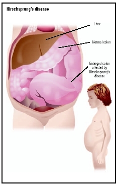
    Hirschsprung’s Disease is a congenital disorder of the large intestine that occurs in about one in five thousand births. Unfortunately, this disease is responsible for twenty-five percent of all intestinal blockages in newborns. Suspected signs and symptoms in a newborn include constipation, abdominal distension, projectile vomiting, poor feeding, and a failure to pass meconium within forty-eight hours after birth. Physiologically, the affected segment of the large intestine narrows because of the absence of local ganglion nerve cells which function to prevent bowel contraction. In long segment HD, more of the large intestine is affected (than in short segment HD) with as much as thirty centimeters of the bowel contracted.  Although the affected area of the large intestine is contracted, the preceding area of the large intestine is enlarged due to the backup of bowel contents which is why patients present with a distended abdomen.

    Pediatricians and pediatric gastroenterologists report that normal breast-fed newborns have approximately four to five bowel movements a day and formula fed newborns will have two to three bowel movements a day. In Hirchsprung’s patients, the number of bowel movements is significantly diminished as a result of the constipation caused by the narrowing of the bowel. At six months of age, most normal newborns have two bowel movements per day; however, this number is typically less for Hirschsprung’s patients of the same age.

    Without proper diagnosis and treatment, Hirschsprung’s patients are at risk for intestinal rupture or perforation that can cause death. Methods of diagnosis include a rectal suction biopsy, or a full thickness biopsy which is generally definitive. Other techniques that may indicate the presence of Hirchsprung’s disease include anorectal manometry and barium enema. There are various surgical techniques available to treat severe Hirschsprung’s disease. Corrective surgery to the large intestine is performed in two phases. The first phase involves a colostomy and is usually performed early in life. Years later, the second surgical phase is performed which is a pull-through procedure where the large intestine is reconnected to the anus. There are several different types of pull-through procedures that are well established, with the transanal pull-through procedure gaining popularity. High fiber diets and frequent enemas help to alleviate symptoms of constipation associated with Hirschsprung’s disease.

    Medical malpractice may arise when there is a failure to diagnose Hirschsprung’s disease that results in death or serious permanent injury. Allegations may include a failure to timely refer the patient to a pediatric gastroenterologist or surgeon, or a failure to order or perform proper diagnostic testing or biopsy.

    For a complete overview on the diagnosis and treatment of Hirschsprung’s disease, please see the 3rd edition of Hirschsprung’s Disease and Allied Disorders by A.M Holschneider and P. Puri which became available in paperback in 2010. For families who have a loved one afflicted with Hirschsprung’s disease, there is a support group called the Hirschsprungs & Motility Disorders Support Network .