Medical Malpractice Attorneys
  • Toll Free 1-800-529-9120
  1. Hypertrophic Pyloric Stenosis

    Posted by Dr. Jack Sacks, Esq.on April 03, 2011

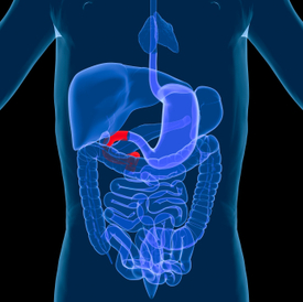
    Hypertrophic pyloric stenosis (HPS) in infants is a narrowing of the pylorus, the lower part of the stomach through which food and other stomach contents pass to enter the small intestine. When an infant has HPS, the muscles in the pylorus have enlarged resulting in narrowing within the pyloric channel preventing food from emptying out of the stomach.

    HPS is a form of gastric outlet obstruction, which means a blockage from the stomach to the intestines. It affects three out of every thousand babies born in the United States. It’s about four times more likely to occur in firstborn male infants and also has been shown to run in families.  HPS occurs more commonly in Caucasian infants than in babies of other ethnic backgrounds.

    Despite numerous hypotheses, the exact cause of HPS is not fully understood. It is believed that these babies are not born with it, but that the progressive thickening of the pylorus occurs after birth. A baby will start to show symptoms when the pylorus is so thickened that the stomach can no longer empty properly.

    The thickening of the pyloric muscles may be a due to a combination of several factors.  The use of erythromycin in the first 2 weeks of life has been associated with HPS, and there is also a connection in babies whose mothers took this antibiotic at the end of pregnancy or during breastfeeding.

    Symptoms of HPS include:

    Projectile vomitinforceful ejection of milk or formula up to several feet away, within 30 minutes after feeding. Vomiting may be mild at first and gradually become more severe. The vomit may sometimes contain blood. Babies are usually not ill-looking or febrile. In the early stage of the disease they remain hungry and suck vigorously after episodes of vomiting.

    Persistent hunger Babies often want to eat soon after vomiting

    Stomach contractions These are noticeable wave-like contractions that move across the upper abdomen (peristalsis) soon after feeding but before vomiting. This is caused by stomach muscles trying to force food past the outlet of the pylorus

    Dehydrationprolonged delay in diagnosis can lead to dehydration. The infant with cry without tears or become lethargic. Diapers need to be changed less often and won’t be as wet as wet as they should be.

    Changes in bowel movementssince food is prevented from reaching the intestines, babies with this condition may be constipated.

    Weight problems– babies will not gain weight, and can sometimes even lose weight.  Parents often report trying several different baby formulas because they (or their physicians) assume vomiting is due to intolerance.

    There are other conditions that can resemble HPS, such as gastroesophageal reflux disease (GERD).  This usually begins at eight weeks of age, with excess spitting up, or reflux, occurring after feedings. However, the majority of infants with GERD do not experience projectile vomiting, and although they may have poor weight gain, they tend to have normal stools.

    In infants with gastroenteritis, which is an inflammation in the digestive tract that can be caused by viral or bacterial infection, their symptoms may also  resemble HPS. Vomiting and dehydration are seen with both conditions; however, infants with gastroenteritis will often have diarrhea with loose, watery, or sometimes bloody stools. Diarrhea usually isn’t seen with HPS.

    Once HPS is diagnosed, early consultation with a surgeon familiar with neonatal care is warranted because treatment is essentially surgical.  This is especially important if the child requires transfer to another facility for surgical care. The American Pediatric Surgical Association offers guidelines for appropriate consultation and transfer of small infants. Good outcome has been shown to depend on the quality of preoperative correction of fluid and electrolyte abnormalities, availability of a pediatric anesthetist, and training level of the surgeon.

    Pyloromyotomy, the surgical procedure in which an incision is made in the longitudinal and circular muscles of the pyloris is the procedure of choice. Laparoscopic pyloromyotomy has a significantly shorter recovery time compared with open pyloromyotomy. However, open pyloromyotomy has a higher efficacy and fewer complications.  Endoscopic pyloromyotomy, another alternative, can be performed as an outpatient procedure. Recently, endoscopic balloon dilatation of HPS after failed pyloromyotomy has been used with greater frequency.


  2. Laparoscopic Cholecystectomy- Minimally Invasive Gallbladder Surgery

    Posted by Dr. Jack Sacks, Esq.on March 10, 2011

    The surgical removal of the gallbladder is called cholecystectomy. Gallbladder problems are usually caused by the presence of gall stones, which are small hard masses consisting of cholesterol or bile salts that form in the gallbladder or in the bile duct.  A problem may arise when one or more gallstones block the flow of bile out of the gallbladder.  This may cause swelling, abdominal pain, vomiting, indigestion, and fever.  If a gallstone blocks the common bile duct (the larger bile duct which drains into the small intestine), jaundice may occur.  Removal of the gallbladder is one of the most commonly performed surgical procedures in the United States.  For the past twenty years, gallbladder surgery has been performed laparoscopically.  The medical name for this procedure is laparoscopic cholecystectomy.

    Laparoscopic surgery is performed withthe assistance of a video camera encased by a long thin tube. During a laparoscopic procedure, small incisions are made and plastic tubes called ports are placed through these incisions. The video camera and small thin instruments are then introduced through the ports, which allow access to the inside of the patient. The camera transmits an image of the organs inside the abdomen onto a television screen, which allows the surgeon to see into the patient’s body and perform the surgery.

    Because laparoscopic cholecystectomy does not require the abdominal muscles to be cut, there is less pain, quicker healing, improved cosmetic results, and fewer complications such as infection and adhesions.  Most patients can be discharged on the same or following day, and can return to work in about a week.  With over twenty years of experience, laparoscopic cholecystectomy should be and is a very safe operation.  The overall complication rate is less than 2% when performed by a properly trained surgeon.

    Complications of laparoscopic cholecystectomy are rare, and can include bleeding, infection, pneumonia, blood clots, or heart problems.  Unintended surgical injury to adjacent structures such as the common bile duct, the first part of the small intestine called the duodenum, or other parts of the small bowel may occur and may require another surgical procedure to repair them.  Bile leakage into the abdomen from damaged bile ducts can cause a painful and potentially dangerous infection. Many cases of minor injury to the common bile duct can be managed non-surgically.  Major injury to the bile duct, however, is a very serious problem and may require corrective surgery. This surgery should be performed by an experienced biliary surgeon

    Because laparoscopic cholecystectomy was considered a new procedure in the late 1980s and 90s, what was called a “learning curve” was created to explain complications that did occur.  By observing the learning curve for laparoscopic skills during those early procedures, and applying what was learned to the training of future surgeons during their residency programs, it was believed future injuries could be avoided. Numerous studies concluded that surgeons truly dedicated and interested in learning new laparoscopic procedures needed to set aside a substantial amount of time to acquire the skills needed. Practice in animate and inanimate models, repeated observation with the opportunity to ask questions and the presence of an instructor during the first ten cases were essential.

    A 2005 article in the journal Surgical Endoscopy entitled “Laparoscopic cholecystectomy after the learning curve: what should we expect?” addressed the issue of the increasing common bile duct (CBD) injuries, which started in the late 1980s.   They did a retrospective analysis of laparoscopic cholecystectomies performed at a single institution from that time to the present, and found that eventually they had decreased the complication rate to zero CBD injuries in 1,674 consecutive procedures.  They concluded that injuries of the CBD can be avoided by performing an extensive dissection and by developing a critical view of the operative field to ensure the patient’s safety.   It appears that unlike the late 1980s and 90s, the 21st century finds laparoscopic cholecystectomy to be a mature and safe surgical procedure.

    Though there are certain acceptable risks associated with any kind of operation, the vast majority of laparoscopic gallbladder patients experience extremely few or no complications and quickly return to normal activities.  It is important to remember that before undergoing any type of surgery, whether laparoscopic or open, one must ask his or her surgeon about his or her training and experience in performing laparoscopic cholecystectomy.  Absent unusual circumstances, life altering complications are no longer acceptable in today’s modern practice of laparoscopic cholecystectomy.


  3. Darvocet and Darvon: Recall Linked to Potentially Serious Heart Problems

    Posted by Dr. Jack Sacks, Esq.on January 07, 2011

    iStock_000011384895XSmall.jpgOn November 19, 2010, at the request of the FDA, certain drug companies producing the painkillers Darvon (Propoxyphene) and Darvocet (Propoxyphene and Acetaminophen) issued a recall of these drugs after a study was done that showed a link to potentially serious or fatal heart arrythmias.  When these drugs are metabolized by the body, they convert to a toxic metabolite that may impair the heart’s transmission of electrical impulses. Darvocet and Darvon are both narcotic analgesics (opioids) that have been prescribed by physicians for patients with mild to moderate pain for over thirty years.  The generic form of the active ingredient is called Propoxyphene and was originally introduced by Eli Lily. It is estimated that over 22 million United States citizens have taken these drugs.

    The above mentioned study by Xanodyne Pharmaceuticals of Newport, KY identified a potential link to an abnormal electrical impulse in the heart called QT Prolongation.  This lengthening of time between the Q wave and the T wave may result in a potentially fatal heart arrythmia called ventricular tachycardia. According to the Federal Drug Abuse Warning Network, Darvocet, Darvon, and generic Propoxyphene are related to over 5% of all drug related deaths between 1987 and 2006. It is anticipated that a multitude of lawsuits will be filed by individuals alleging injuries such as heart arrthymia, heart attack, stroke, and suicide.


  4. Accutane and the Potential for Increased Risk of Crohn’s Disease and Ulcerative Colitis

    Posted by Dr. Jack Sacks, Esq.on December 31, 2010

    Accutane has been prescribed since the 1980’s to treat severe acne.  Since then, thousands of patients have reported serious side effects that include gastrointestinal disorders. The American Journal of Gastroenterology reported the connection in 2006, specifically noting the development of the inflammatory bowel diseases Crohn’s and ulcerative colitis in some Accutane users.

    iStock_000012888068XSmall.jpg


    Crohn’s disease usually causes ulcerations and bowel wall swelling in areas throughout the length of the small and large intestine.  Crohn’s disease can spare the rectum, but often causes inflammation or infection with drainage around the rectum.  Ulcerative colitis usually causes ulcers in the lower part of the large intestine, often starting at the rectum.  However, it can involve the entire large intestine and extend into the small intestine (see www.familydoctor.org).

    The FDA requested that Hoffman-La Roche change their label for Accutane to warn that the drug has been associated with the development of inflammatory bowel disease in patients without a prior history of intestinal disorders.  These inflammatory bowel diseases have been reported to persist after Accutane treatment has been stopped. In 2009, Swiss based Roche Holding recalled Accutane from the market due to evidence that the drug was linked to the potential development of inflammatory bowel disease.


  5. Hirschsprung’s Disease and Medical Malpractice

    Posted by Dr. Jack Sacks, Esq.on December 29, 2010

    gech_0001_0002_0_img0134.jpg

    Hirschsprung’s Disease is a congenital disorder of the large intestine that occurs in about one in five thousand births. Unfortunately, this disease is responsible for twenty-five percent of all intestinal blockages in newborns. Suspected signs and symptoms in a newborn include constipation, abdominal distension, projectile vomiting, poor feeding, and a failure to pass meconium within forty-eight hours after birth. Physiologically, the affected segment of the large intestine narrows because of the absence of local ganglion nerve cells which function to prevent bowel contraction. In long segment HD, more of the large intestine is affected (than in short segment HD) with as much as thirty centimeters of the bowel contracted.  Although the affected area of the large intestine is contracted, the preceding area of the large intestine is enlarged due to the backup of bowel contents which is why patients present with a distended abdomen.

    Pediatricians and pediatric gastroenterologists report that normal breast-fed newborns have approximately four to five bowel movements a day and formula fed newborns will have two to three bowel movements a day. In Hirchsprung’s patients, the number of bowel movements is significantly diminished as a result of the constipation caused by the narrowing of the bowel. At six months of age, most normal newborns have two bowel movements per day; however, this number is typically less for Hirschsprung’s patients of the same age.

    Without proper diagnosis and treatment, Hirschsprung’s patients are at risk for intestinal rupture or perforation that can cause death. Methods of diagnosis include a rectal suction biopsy, or a full thickness biopsy which is generally definitive. Other techniques that may indicate the presence of Hirchsprung’s disease include anorectal manometry and barium enema. There are various surgical techniques available to treat severe Hirschsprung’s disease. Corrective surgery to the large intestine is performed in two phases. The first phase involves a colostomy and is usually performed early in life. Years later, the second surgical phase is performed which is a pull-through procedure where the large intestine is reconnected to the anus. There are several different types of pull-through procedures that are well established, with the transanal pull-through procedure gaining popularity. High fiber diets and frequent enemas help to alleviate symptoms of constipation associated with Hirschsprung’s disease.

    Medical malpractice may arise when there is a failure to diagnose Hirschsprung’s disease that results in death or serious permanent injury. Allegations may include a failure to timely refer the patient to a pediatric gastroenterologist or surgeon, or a failure to order or perform proper diagnostic testing or biopsy.

    For a complete overview on the diagnosis and treatment of Hirschsprung’s disease, please see the 3rd edition of Hirschsprung’s Disease and Allied Disorders by A.M Holschneider and P. Puri which became available in paperback in 2010. For families who have a loved one afflicted with Hirschsprung’s disease, there is a support group called the Hirschsprungs & Motility Disorders Support Network .